Pretreatment technique and bioengineered patch for improved stem cell healing of heart failure
Researchers from The Ohio State University Wexner Medical Center (OH, USA) are investigating two methods of maximizing the healing potential of stem cells by helping them overcome the inhospitable conditions of the damaged heart.
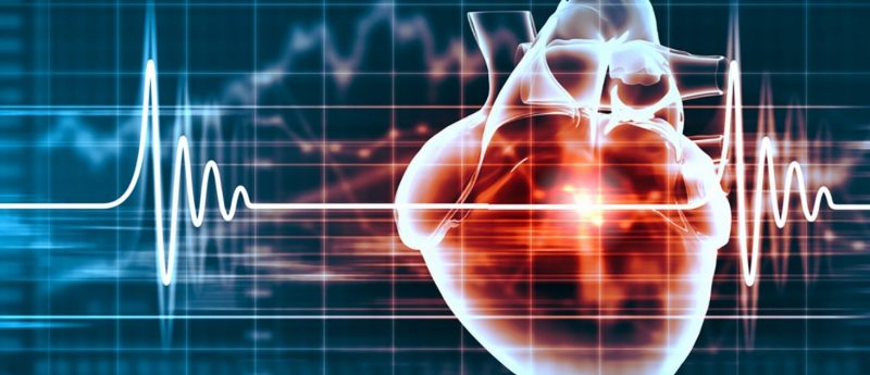
Heart disease is the world’s leading killer, causing the deaths of more than 7.5 million individuals annually, and leaving millions more dependent on medicines and devices to survive. Although many different types of stem cells have been administered in many ways to try and repair or regenerate the damage caused by heart disease, results have not been consistent.
“We’ve seen a lot of tantalizing data over the years, but also a lot of variability in outcomes. Some people have small gains, while others gain nothing,” explains Dr Mahmood Khan, a scientist at the Davis Heart and Lung Research Institute at The Ohio State University Wexner Medical Center (OH, USA). “We still don’t know exactly how stem cells work to heal the heart. But we do know that 90% of stem cells die or disappear within the first 4 days of transplant, so their potential is probably not being fully realized.”
Khan alongside Dr Mark Angelos, Vice Chair of Research in the Department of Emergency Medicine at Ohio State’s College of Medicine, decided to see if they can improve success of stem cell transplants by making the heart microenvironment more hospitable. “A damaged heart has areas of poor oxygenation and blood flow. It is not a friendly place for stem cells to thrive,” stated Angelos. “By developing techniques that improve stem cell delivery and survival, we’re hopeful that the therapy will be able to yield more consistent and greater benefits.”
It is believed that the most effective time to administer stem cell therapies after a myocardial infarction is within hours to days, as this is when oxygen-deprived cells begin apoptosis, which generates the scar tissue that causes permanent heart failure. “It’s the probably the best and worst time to deliver stem cell therapies. We are literally sending them into battlefield where there is minimal oxygen and genes are telling cells to self-destruct,” Khan commented.
As miRNAs regulate 30% of all mammalian protein-encoding genes, they may be a therapeutic target for many diseases. In the case of heart disease, miR-133a appears to regulate fibrosis and cardiac remodeling, and is found to be decreased in people who have suffered a heart attack. Therefore, the research team hypothesized that increasing miR-133a levels in cultured stem cells may help them to survive in the heart, and bioengineered a molecule to stimulate mesenchymal stem cells (MSCs) to generate miRNA-133a. When transplanted into an animal model of cardiac ischemia, the cells demonstrated improved survival over non-treated MSCs.
Angelos commented: “We found that the pretreated MSCs did a better job at decreasing the global damage to the heart, along with improvement in the left-ventricular wall thickness compared to the untreated MSCs… MSCs are a commonly used cell type in current heart failure studies, so our findings are definitely relevant to that work.”
The team also decided to investigate how to help stem cells fully integrate and function alongside the existing heart tissue. “The heart is a constantly moving, connected matrix of muscle fibers working together to make the heart pump in sync,” explains Dr Angelos. “Transplanted stem cells may not align with native tissue, potentially disrupting or attenuating signals that keep a steady heartbeat. There’s evidence that this could contribute to arrhythmias.”
To create a more secure environment for stem cell integration, Drs Khan and Angelos used a biodegradeable nanofiber ‘patch’ seeded with human inducible pluripotent stem cell-derived cardiomyocytes (hiPSC-CMs). hiPSC-CMs were seeded onto the aligned nanofiber patch and a standard culture plate, and compared for calcium signaling and synchronous beating. Within 2 weeks, both demonstrated spontaneous beating like a miniature heart, but the linear grain of the nanofiber formed an aligned pattern of cells that more greatly resembled and functioned healthy heart tissue. “Next, we hope to use what we’ve learned from this study to develop a thicker, multi-layer patch that could help restore thin and weakened heart walls,” stated Khan.
Drs Khan and Angelos believe that their miRNA pretreatment and cardiac patch methods hold great potential for improving outcomes in heart failure patients, by providing stem cells with a survival boost and a protective structure.