Getting to know EMT: Re-epithelialization during Wound Healing
Considerations when studying epithelial-to-mesenchymal transition in the context of wound healing.
Getting to Know EMT: Re-epithelialization During Wound Healing
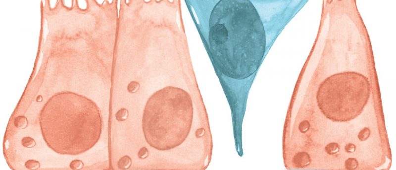
Epithelial-to-Mesenchymal Transition (EMT) refers to the process where cells within stationary epithelial tissue change into a more migratory, mesenchymal-like phenotype. In the realm of regenerative medicine, EMT contributes to the process of wound healing, providing a critical mechanism for the re-epithelialization of tissue following injury. Understanding the biology that underlies EMT is important for developing therapeutics that facilitate wound healing and tissue regeneration, including treatments that prevent organ fibrosis and tissue scarring, an event that can occur if EMT persists following the attenuation of injury-induced inflammation.
This article discusses the basics of EMT and some techniques and resources for studying it.
Molecular Mechanisms of EMT
MT is a biological process by which differentiated epithelial cells lose epithelial characteristics and acquire a migratory, mesenchymal phenotype. While our understanding of the transitory phenotype of cells undergoing EMT is evolving, much is known about its induction and completion. Typically, epithelial cells display apical-basal polarity and adhere tightly to each other via tight and adherens junctions near the apical membrane and desmosomes in the basolateral membrane. The morphological changes that occur during EMT are induced by signal transduction pathways that reduce E-Cadherin expression, drive the disassembly of intercellular adhesion complexes, and promote Actin stress fiber and focal adhesion formation. These types of cellular changes result in the phenotypic transition to an elongated, mesenchymal cell that expresses extracellular matrix remodeling enzymes and has an increased capacity for migration and invasion. EMT and the reverse process, mesenchymal to epithelial transition (MET), are thought to be controlled by local cues within distinct microenvironments.
For a more detailed description of EMT mechanisms, please view or download our Poster on Epithelial to Mesenchymal Transition.
Methods to Study EMT
 Cell Characterization: A critical step to studying EMT is being able to clearly distinguish epithelial and mesenchymal phenotypes. Antibodies are readily available against cell surface markers and transcription factors specific to epithelial and mesenchymal phenotypes. A great reference for these markers and corresponding antibodies can be found at Novus Biologicals website. To make it more simple, one-step simultaneous immunofluorescence staining of human epithelial and mesenchymal markers can be performed with the aptly named Human EMT 3-Color Immunocytochemistry Kit from R&D Systems, which features a combination of fluorphore-conjugated antibodies against E-Cadherin (epithelial marker), Snail and Vimentin (mesenchymal markers).
EMT Induction: Cell culture models of EMT are employed to understand the biology of EMT and for identifying and evaluating novel prognostic markers and therapeutics for fibrotic diseases, wound healing, and metastasis. The ability to induce EMT in vitro typically requires TGF-beta stimulation or labor-intensive genetic modification. While TGF-beta stimulation is the most common method used to induce EMT, it is not always sufficient. To more reliably induce EMT, specialized cocktails of recombinant proteins and neutralizing antibodies can be utilized. One such cocktail is the StemXVivo® EMT Induction Media Supplement from R&D Systems. This supplement, which contains an optimized cocktail of proteins and antibodies, is designed for straightforward and quick induction of EMT.
 Functional Assays: A key aspect of EMT as it relates to wound healing is the migration of mesenchymal-like cells into the injury site of injury. Chemotactic assays are a standardized and effective way to monitor the migration of mesenchymal stem cells after EMT induction. These assays enable researchers to assess the efficiency of EMT induction as well as investigate the mechanisms underlying re-epithelialization through migration. In the data example to the right, EMT Induction Media was used to trigger EMT in a number of cancer cell lines and the efficiency of induction was quantified using cell invasion and migration assays.
Comprehensive Tools for EMT
For a more comprehensive report on tools and techniques used for EMT, please view our video “Induction and Analysis of Epithelial to Mesenchymal Transition,” published in the Journal of Visualized Experiments. Or you can visit the EMT Frequently-Asked-Questions webpage by Novus Biologicals.