Forks in the road: cell signaling and gene expression in the determination of mesodermal cell fates
Study by researchers at Stanford University School of Medicine (CA, USA) identifies the signals required to support quicker and more efficient differentiation of human embryonic stem cells into specialized tissues that could be used in the clinic.
Differentiation of cell types from embryonic cells is a complex process involving specific cell signaling to determine the fate of each cell, and culturing pure populations within days rather than weeks to months would increase their clinical utility. Therefore, researchers based at Stanford University School of Medicine (CA, USA) have been investigating how to map out particular cell signals that are important in directing the development of human embryonic stem cells to quickly form pure populations of a number of transplantable human tissue progenitors, including bone, heart muscle and cartilage cells.
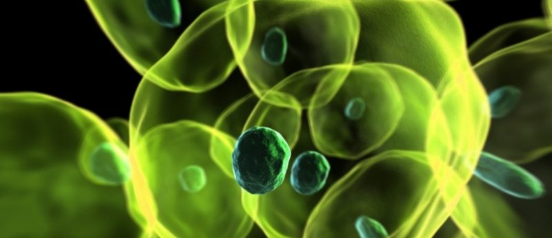
Although little of the embryonic developmental process is known due to law restricting culturing of human embryos longer than 12 days is many countries, it is known that the embryo consists of three main components in its early stages: the ectoderm, endoderm and the mesoderm, which then generate the more specific cell types. In particular, the mesoderm produces cardiac and skeletal muscle, connective tissue, bone, blood vessels, blood cells, cartilage and portions of the kidneys and skin.
The Stanford researchers were looking specifically at the signals required to give rise to mesodermally derived cells. They took a human embryo cell line and encouraged the cells to form the primitive streak, then used signaling molecules, such as WNT, BMP and Hedgehog, to specialize the cells. They found that cells will normally progress down two pathways, deciding between two options.
Cells in the primitive streak can either become endoderm or two types of mesoderm, and inhibiting certain signaling molecules forced the cells to take a particular path. In this case, inhibiting TGF-b drove cells to be mesodermal. When WNT was added and BMP was blocked, the cells chose one of the two types of mesoderm, and conversely would chose the other type when WNT was blocked and BMP added. Loh and Chen guided the cells to become bone cell precursors, which were transplanted into laboratory mice and became human bone, beating heart muscle cells and ten other mesoderm-derived cell lineages.
Loh and Chen also carried out single-cell RNA sequencing at each developmental stage to look at the purity of the cell populations, and use the gene expression profile to report on transient states that can be associated with development from a precursor to the more specialized cell. Similar to a process already seen in other animals, the researchers saw a brief rise in gene expression at the point just before the human embryo splits to segments that will become the head, trunk and limbs.
Loh commented that, “The segmentation of the embryo is a fundamental step in human development. Now we can see that, evolutionarily, it’s a very conserved process.” This information on the segmentation process could be a way to further understand how congential birth defects occur.
Finally, Loh explained that: “Next, we’d like to show that these different human progenitor cells can regenerate their respective tissues and perhaps even ameliorate disease in animal models.”
— Written by Emily Hargrave
Source: KM Loh, Chen A, Koh PW et al. Mapping the pairwise choices leading from pluripotency to human bone, heart, and other mesoderm cell types. Cell doi:10.1016/j.cell.2016.06.011 (2016) (Epub ahead of print); http://med.stanford.edu/news/all-news/2016/07/researchers-coax-human-stem-cells-to-rapidly-generate-bone.html