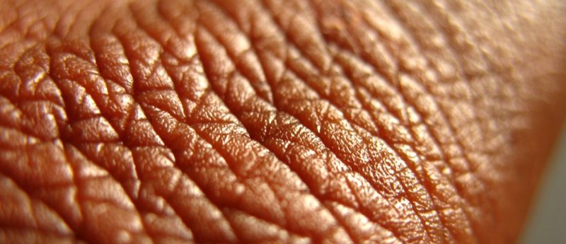
hESC/iPSC-based 3D skin models — customizing therapy of atopic dermatitis
How iPSC technology can be utilized to build advanced models for atopic dermatitis research and drug testing.
Matthew Donne1 and Dusko Ilic2
1VitroLabs Inc., San Francisco, CA 94104, USA
2Division of Women’s Health, Faculty of Science and Medicine, King’s College London, London, UK.
Correspondence: [email protected]
Atopic dermatitis (AD) or eczema is a devastating inflammatory, chronically relapsing, noncontagious and extremely pruritic skin disease. AD affects an estimated 15—30% of children and 2—10% of adults worldwide. Of those, 20% have moderate-to-severe forms of the disease [1]. Whereas much research in AD has focused upon the adaptive immune response, new data suggest that allergic sensitization may occur secondary to impairment of epidermal barrier function — the outermost layer of the epidermis, the stratum corneum, serves as the protective barrier against dehydration and penetration of exogenous agents [2,3]. The FLG gene encodes a key protein that facilitates terminal differentiation of the epidermis and formation of the skin barrier. It has been found that the loss-of-function genetic variants in the FLG gene are very strong AD predisposing factors [2,4].
Current management of AD
Skin diseases are very common around the world, affecting 30—70% of the population across all age groups and were the 4th leading cause of nonfatal burden expressed as “years lost due to disability (YLD)” in 2010 [5]. On a scale from 0 (perfect health) to 1 (equivalent to death), average disability weight for AD is 0.038, the second highest among skin diseases (the highest is skin cancer). Strikingly different severity and extent of disease seen in AD patients, and lack of generalized treatment, highlight the need of personalized approach to patient care.
The management of AD follows the individual symptomatic variability of disease [6-8]. Basic therapy is focused on hydrating topical treatment with various emollients and avoidance of specific and unspecific provocation factors. Anti-inflammatory treatment with topical antihistamines [9], glucocorticoids and calcineurin inhibitors remain the standard procedure for more severe cases [6-8]. Oral cyclosporin is approved for systemic treatment, but classic immunosuppressants azathioprine and methotrexate have also shown efficacy in clinical trials. Potentially detrimental adverse effects limit the use of these drugs.
Up to 50% of AD patients have abandoned traditional healthcare providers because they have nothing new to offer [10]. Recently published phase I and II studies suggest that dupilumab, a monoclonal antibody that targets a subunit of the interleukin-4 (IL-4) and IL-13 receptors, has the potential to become the first systemic therapy for atopic dermatitis to be approved by the US FDA [11]. Dupilumab, developed by Regeneron and Sanofi, has previously shown efficacy in a Phase II trial for asthma [12]. However, although the results seem to be promising, the therapy is not targeting the underlying cause — permeability barrier defect.
High social impact of AD
Current clinical care protocols are limited only to ameliorating the symptoms. A recent study on the psychological burden of skin diseases among dermatological outpatients in 13 European countries found that 8.0—15.1% of AD patients were clinically depressed and 16.7—21.0% displayed anxiety [13]. In a study from Germany, 16% of AD patients had suicidal ideation compared with 1% of the controls [14]. In 2007, a study quantified the incremental direct medical and indirect work-loss costs associated with patients diagnosed with AD utilizing a de-identified administrative claims database, which comprised 5.1 million covered beneficiaries from 31 Fortune 500 self-insured employers between 1998 and 2005 [15].
The total incremental cost per patient per month for the AD group was US$83 (direct: US$52, P<0.001; indirect: US$31, P<0.001). Employer-payers experience a significant annual cost burden of US$ 991 per patient attributable to AD. Employee disability and increased number of sick days account for 38% of the cost burden. Since nearly 10% of the European Union (EU) population of 500 million carries this mutation in FLG and have reported AD-linked symptoms [2], potential savings are enormous. Cutting costs for only 1% by introducing personalized care of AD patients would save the EU nearly US$500 million annually.
Limitations of the current AD models
Currently available animal models of AD include mice, guinea pigs and dogs. However, none of them can be used to correlate extent of the symptoms and genetic polymorphism of AD patients. In addition to ethical problems surrounding the use of animals in research, complexity of the disease does not allow us to dissect in vivo interdependence permeability barrier defect and immune response to enhanced penetration of external allergens.
In vitro models are invaluable for exploring the etiopathogenesis of AD and validating the safety and effectiveness of drug candidates [16, 17]. Various models based on human cell and tissue cultures have been developed [18], although all of them are relatively simplistic. Thus, there is a pressure for development of more sophisticated relevant in vitro models to validate new drug candidates for the treatment of AD.
hESC/iPSC-based human skin equivalents are indispensable tools for the future of AD research
Human epidermal equivalents (HEEs) that parallel epidermal properties are essential for studying barrier development and function. In spite of advances in HEE engineering, generating functional permeability barrier in vitro was rarely successful [19, 20]. The studies have been further limited by the fact that only a limited number of HEE can be generated from one sample of epidermis and the primary keratinocytes generated from this sample may contain previously unidentified polymorphisms in genes involved in barrier development. The addition of fibroblasts or de-epidermized dermis further contributes to the batch-to-batch variability. The quantitative and qualitative changes in epidermal barrier and activation of immune cells are highly codependent. Therefore, a robust AD-HEE model should also contain cells of the immune system.
We recently developed a HEE model that can be produced in an unlimited number of genetically identical units, utilizing keratinocytes differentiated from human embryonic stem cells (hESCs)/induced pluripotent stem cells (iPSCs), primary cells that are capable of infinite proliferation and whose genetic footprint can be fully characterized [21]. Developing this model further — engineering HSE containing both epidermal and dermal elements — might be the answer to the need of the field of AD.
Beside keratinocytes, epidermis should contain Langerhans cells. They are involved in immunological T-cell responses and implicated in the etiopathogenesis underlying a variety of allergic and inflammatory skin diseases. They, together with mast cells, have a critical role in the development of AD [22, 23]. hESC/iPSC-based HSE models would allow the utilization of cells with different mutations in FLG for dissecting molecular events in AD as well as screening and matching AD patients with treatments that are more likely to be effective and cause fewer side effects.
The strategy would be generating AD-specific iPSC lines from donors harboring each of the most common mutations in FLG (FLG-iPSC). To minimize the possibility of data misinterpretation due to genetic and epigenetic footprints of the cell lines, two sets of complementary stem cell lines should be generated with help of the CRISPR-Cas9 gene editing system [24, 25].
To obtain “wild-type” control of FLG-iPSC, FLG mutation can be reverted and, by the same token, the FLG mutation can be introduced into normal healthy hESC lines. Therefore, each of the AD-linked mutations in FLG would have iPSC (reprogrammed, edited) and hESC (not reprogrammed, not edited) “wild-type” controls and their mutant iPSC (reprogrammed, not edited) and hESC (not reprogrammed, edited) counterparts. Building such robust model system will provide the medical technology sector with a major competitive advantage and has a strong potential to move forward the field of iPSC-derived “disease models” for screening of compounds.
As the field of systems dermatology prepares to expand its efforts [26], this is an ideal time to introduce a novel integrated theoretical and experimental systems biology approach, to be combined with stem cell technology and novel gene editing techniques to advance our understanding of the systems-level dynamics of AD towards designing novel AD therapies.
Financial & competing interest disclosure
- Matthew Donne has no competing interests. Dusko Ilic has a grant from LEO Pharma to develop the model described in the article.
References
- http://www.merckmanuals.com/professional/dermatologic_disorders/dermatitis/atopic_dermatitis.html [Accessed 03/03/2017].
- McGrath JA, Uitto J. The filaggrin story: novel insights into skin-barrier function and disease. Trends Mol. Med. 14(1), 20-27 (2008).
- Scharschmidt TC, Man MQ, Hatano Y et al. Filaggrin deficiency confers a paracellular barrier abnormality that reduces inflammatory thresholds to irritants and haptens. J. Allergy Clin. Immunol. 124(3), 496-506 (2009).
- Palmer CN, Irvine AD, Terron-Kwiatkowski A et al. Common loss-of-function variants of the epidermal barrier protein filaggrin are a major predisposing factor for atopic dermatitis. Nat. Genet. 38(4), 441-446 (2006).
- Hay RJ, Johns NE, Williams HC et al. The global burden of skin disease in 2010: An analysis of the prevalence and impact of skin conditions. J. Invest. Dermatol. 134(6), 1527-1534 (2014).
- Ring J, Alomar A, Bieber T et al. Guidelines for treatment of atopic eczema (atopic dermatitis) Part I. J. Eur. Acad. Dermatol. Venereol. 26(8), 1045-1060 (2012).
- Ring J, Alomar A, Bieber T et al. Guidelines for treatment of atopic eczema (atopic dermatitis) Part II. J. Eur. Acad. Dermatol. Venereol. 26(9), 1176-1193.
- Wollenberg A, Oranje A, Deleuran M et al. ETFAD/EADV Eczema task force 2015 position paper on diagnosis and treatment of atopic dermatitis in adult and paediatric patients. J. Eur. Acad. Dermatol. Venereol. 30(5), 729-747 (2016).
- Lin TK, Man MQ, Santiago JL et al. Topical antihistamines display potent anti-inflammatory activity linked in part to enhanced permeability barrier function. J. Invest. Dermatol. 133(2), 469-478 (2013).
- Cully M. Trial watch: Atopic dermatitis therapy breakthrough on the horizon? Nat. Rev. Drug Discov. 13(9), 645 (2014).
- Beck LA, Thaçi D, Hamilton JD et al. Dupilumab treatment in adults with moderate-to-severe atopic dermatitis. N. Engl. J. Med. 371(2), 130-139 (2014).
- Wenzel S, Ford L, Pearlman D et al. Dupilumab in persistent asthma with elevated eosinophil level. N. Engl. J. Med. 368(26), 2455-2466 (2013).
- Dalgard FJ, Gieler U, Tomas-Aragones L et al. The psychological burden of skin diseases: A cross-sectional multicenter study among dermatological out-patients in 13 European countries. J. Invest. Dermat. 135(4), 984-991 (2015).
- Dieris-Hirche J, Gieler U, Kupfer JP, Milch WE. Suicidal ideation, anxiety and depression in adult patients with AD. Hautarzt. 60(8), 641-646 (2009).
- Fowler JF, Duh MS, Rovba L et al. The direct and indirect cost burden of atopic dermatitis: an employer-payer perspective. Manag. Care Interface 20(10), 26-32 (2007).
- Thakoersing VS, Gooris GS, Mulder A, Rietveld M, El Ghalbzouri A, Bouwstra JA. Unraveling barrier properties of three different in-house HSEs. Tissue Eng. Part C Methods. 18(1), 1-11 (2012).
- Thakoersing VS, van Smeden J, Boiten WA et al. Modulation of stratum corneum lipid composition and organization of human skin equivalents by specific medium supplements. Exp. Dermatol. 24(9), 669-674 (2015).
- Castex-Rizzi N, Galliano MF, Aries MF et al. In vitro approaches to pharmacological screening in the field of atopic dermatitis. Br. J. Dermatol. 170 Suppl 1, 12-18 (2014).
- Bouwstra JA, Groenink HW, Kempenaar JA, Romeijn SG, Ponec M. The water distribution and natural moisturizer factor in human skin equivalents grown at various relative humidities. J. Invest. Dermatol. 128(2), 378-388 (2008).
- Sun R, Celli A, Crumrine D et al. Lowered humidity produces HEE with enhanced barrier properties. Tissue Eng. Part C Methods. 21(1), 15-22 (2015).
- Petrova A, Celli A, Jacquet L et al. 3D in vitro model of a functional epidermal permeability barrier from hESC and iPSC. Stem Cell Reports. 2(5), 675-689 (2014).
- Artuc M, Steckelings UM, Grützkau A, Smorodchenko A, Henz BM. A long-term coculture model for the study of mast cell-keratinocyte interactions. J. Invest. Dermatol. 119(2), 411-415 (2002).
- Elentner A, Finke D, Schmuth M, Chappaz S et al. Langerhans cells are critical in the development of atopic dermatitis-like inflammation and symptoms in mice. J. Cell Mol. Med. 13(8B), 2658-2672 (2009).
- Jinek M, Chylinski K, Fonfara I, Hauer M, Doudna JA, Charpentier E. A programmable dual-RNA-guided DNA endonuclease in adaptive bacterial immunity. Science. 337(6096), 816-21 (2012).
- Smurnyy Y, Cai M, Wu H et al. DNA sequencing and CRISPR-Cas9 gene editing for target validation in mammalian cells. Nat. Chem. Biol. 10(8):623-625 (2014).
- Reynolds NJ. One hundred and twenty-five years and counting: into an era of systems dermatology. Br. J. Dermatol. 171(6), 1279-1281 (2014).