Step forward in human tissue printing
Scientists demonstrate new technique and prove feasibility of printing living tissue structures for implantation
A recent report has announced printing of ear, bone and muscle structures using a custom-designed 3D printer. When implanted in animals, the living tissue structures matured into functional tissue and developed a system of blood vessels. The authors of the study suggest that these early results indicate that the structures have the right size, strength and function for use in humans.
This novel tissue and organ printer is an important advance in our quest to make replacement tissue for patients,” explained senior author Anthony Atala (director of the Wake Forest Institute for Regenerative Medicine, NC, USA). “It can fabricate stable, human-scale tissue of any shape. With further development, this technology could potentially be used to print living tissue and organ structures for surgical implantation.”
Using funding from the US Armed Forces Institute of Regenerative Medicine, established to advance the translation of regenerative medicine for battlefield injuries, Atala’s team hope to one day implant bioprinted muscle, cartilage and bone into patients.
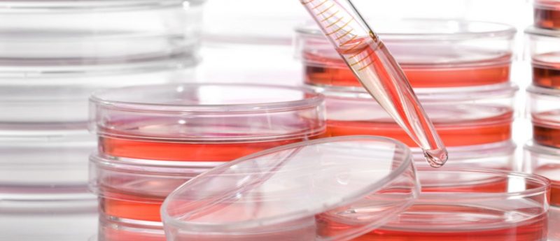
It is widely hoped that 3D printing will be revolutionary as a method for replicating the body’s complex tissues and organs; however, current printer methods based on jetting, extrusion and laser-induced forward transfer cannot produce structures with sufficient size or strength for implantation in the body. The Integrated Tissue and Organ Printing System, developed over a 10-year period by scientists at the Institute for Regenerative Medicine, aims to overcome these challenges. The system deposits both biodegradable, plastic-like materials to form the tissue shape and water-based gels that contain the cells. A temporary strong outer structure is also formed.
At this stage, a major challenge of tissue engineering is ensuring that implanted structures live long enough in the body. The new technique discussed in this paper addresses this in two ways: the water-based ink that holds the cells was optimized to promote cell health and growth; and a lattice of microchannels was printed throughout the structures. These channels allow nutrients and oxygen from the body to diffuse into the structure and keep them alive while they develop a system of blood vessels.
In this investigation, researchers observed an ear structure of over 35 mm to survive and show signs of vascularization at one and two months after implantation, a notable improvement on previous findings which indicated that tissue structures without ready-made blood vessels must be smaller than 200 microns for cells to survive. “Our results indicate that the bio-ink combination we used, combined with the micro-channels, provides the right environment to keep the cells alive and to support cell and tissue growth,” explained Atala.
Following several proof-of-concept experiments demonstrating the method’s capabilities with external ear structures, muscle tissue and jaw bone fragments, studies are ongoing to measure longer-term outcomes.
— Written by Hannah Wilson