Knee meniscus tissue engineering
A brief introduction to the challenge of knee meniscus tissue engineering, and the current techniques and technologies under investigation
The menisci
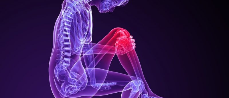
The menisci are two crescent-shaped fibro cartilaginous structures that are found between femur and tibia (Fig. 1). Menisci act as a shock absorber which helps to dissipate the forces in the knee caused by all activities and spread the load of the body’s weight over a greater area. The most common causes of a meniscus tear are traumatic injury (often seen in defense personnel and sportsmen) and degenerative processes (seen in older patients who have more brittle menisci) [1].
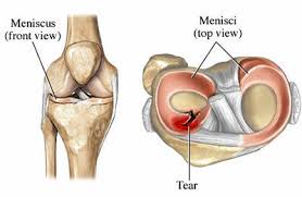
Fig.1 : Knee Meniscus
This damaged meniscus often takes more time to heal or remain unhealed due to the avascular region present in inner one third of meniscus. These variations as well as the non-healing ability of meniscus need meniscectomy (partial or complete). Such removal of meniscus through meniscectomy causes various problems to the patients including osteoarthritis, bone degeneration, excess pain etc. Hence in order to overcome these problems, tissue engineering using various scaffolds has been tried as a new treatment modality for meniscus tissue regeneration [1].
Tissue engineering approaches for repairing the menisci
Tissue engineering approaches combining biomaterials, cells, and growth factors have been investigated for the treatment of meniscal defects in vitro and in vivo using various animal model. The commonly used biomolecules like biotin, proline, glucose and chondroitin sulphate [2] are cost effective and affordable as compared to other expensive growth factors such as TGF-β [3], BMP [4], etc. used for enhancement of cell growth and proliferation. Various polymers like PCL [5], polyurethane [6], silk [7], egg shell membrane [8] etc., have been studied for preparation of scaffold using different methods such as electrospinning (nanofibers) [5], lyophilization (3D sponges) [9], 3D printing [10] etc.
Recently, three dimensional (3D) printing has been investigated for the fabrication of scaffolds with various shapes and structure. Development of 3D scaffolds that can replace the natural extracellular matrix will allow the diffusion of nutrient, metabolites and soluble factors until the seeded cells can produce a new matrix and regenerate the tissue structures. This may yield an in vitro regenerated meniscus tissue with functional properties which will be similar to native human meniscus cartilage. Thus tissue engineering approach not only minimizes the risk for developing knee osteoarthritis but also offers a solution for knee meniscus problems.
Our team at PSG Institute of Advanced Studies has been working in the area of meniscus tissue engineering for last five years. Scaffold based meniscal regeneration and scaffold free meniscal development are two different modalities tried to regenerate meniscus. In our previous study, we have developed a scaffold free meniscal tissue using high density pellet culturing (HDPC) which has perfect distribution of collagen fibers as found in native human meniscus [11]. We have also reported that addition of a unique combination of biomolecule (UCM) has impact on reducing the time taken for proliferation of primary human meniscal cells [2]. We also developed functionalized (using biomolecules) scaffolds like sponge, film and nanofiber using natural and synthetic polymers like PCL [6], silk [7] and egg shell membrane [8]. These scaffolds cytocompatibility, cell adhesion and proliferation studies were carried out using primary human meniscus cells isolated from surgical debris. The main challenge in meniscus tissue engineering is mimicking the structure and functional mechanical properties of this cartilage. However, currently no such biomaterials are there that can simulate the tensile, compressive and frictional properties of meniscus cartilage [12]. Our team is working on this strategy and we hope that this can be achieved in near future.
References
- Chambers MC, El-Amin SF. Tissue engineering of the meniscus: scaffolds for meniscus repair and replacement. Musculoskeletal Regeneration 2, e998 (2015).
- Pillai MM, Elakkiya V, Gopinathan J et al. A combination of biomolecules enhances expression of E-cadherin and peroxisome proliferator-activated receptor gene leading to increased cell proliferation in primary human meniscal cells: an in vitro study. Cytotechnology 28, 1—5 (2015).
- Demoor M, Ollitrault D, Gomez-Leduc T et al. Cartilage tissue engineering: molecular control of chondrocyte differentiation for proper cartilage matrix reconstruction. Biochimica et Biophysica Acta 1840(8), 2414—2440 (2014).
- Perrier-Groult E, Pasdeloup M, Malbouyres M, Galéra P, Mallein-Gerin F. Control of collagen production in mouse chondrocytes by using a combination of bone morphogenetic protein-2 and small interfering RNA targeting Col1a1 for hydrogel-based tissue-engineered cartilage. Tissue Eng. Part C Methods 19(8), 652—664 (2013).
- Gopinathan J, Mano S, Elakkiya V, Pillai MM, Sahanand KS, Rai BD, Selvakumar R, Bhattacharyya A. Biomolecule incorporated poly-ε-caprolactone nanofibrous scaffolds for enhanced human meniscal cell attachment and proliferation. RSC Advances 5(90), 73552— 73561 (2015).
- Verdonk R, Verdonk P, Huysse W, Forsyth R, Heinrichs EL. Tissue ingrowth after implantation of a novel, biodegradable polyurethane scaffold for treatment of partial meniscal lesions. Am. J. Sports Med. 39(4), 774—782 (2011).
- Varkey A, Venugopal E, Sugumaran P et al. Impact of silk fibroin-based scaffold structures on human osteoblast MG63 cell attachment and proliferation. Int.. Nanomedicine. 10(Suppl. 1), 43 (2015).
- Pillai MM, Akshaya TR, Elakkiya V et al. Egg shell membrane — a potential natural scaffold for human meniscal tissue engineering: an in vitro study. RSC Advances 5(93), 76019—76025 (2015).
- Hoyer B, Bernhardt A, Lode A et al. Jellyfish collagen scaffolds for cartilage tissue engineering. Acta Biomater. 10(2), 883-92.z (2104).
- Lee CH, Rodeo SA, Fortier LA, Lu C, Erisken C, Mao JJ. Protein-releasing polymeric scaffolds induce fibrochondrocytic differentiation of endogenous cells for knee meniscus regeneration in sheep. Sci. Transl. Med. 6(266), 266ra171 (2014).
- Pillai MM, Elakkiya V, Gopinathan J et al. High density pellet culture for human knee meniscus tissue formation using a unique combination of medium. Regen. Med. 10(7s), 58 (2015).
- Huey DJ, Hu JC, Athanasiou KA. Unlike bone, cartilage regeneration remains elusive. Science 16, 338(6109), 917—921 (2012).