A biomedical engineer’s role in multidisciplinary pre-surgical planning and practice at the point-of-care


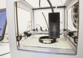
Reconstruction of the face and jaw remains one of the primary uses of 3D planning and printing at Mayo Clinic in Rochester (MN, USA). In this editorial, Biomedical Engineer, Amy Alexander, describes how biomedical engineering influences and compliments pre-operative preparation with 3D printing.
Biography

Amy Alexander is a Senior Biomedical Engineer in the Mayo Clinic Department of Radiology’s Anatomic Modeling Lab in Rochester (MN, USA). In her role, Alexander converts 2D radiological images into 3D models and patient-specific surgical cutting guides. These life-size, patient-specific models and guides help surgeons from different specialties prepare for complex procedures. Additionally, these 3D prints form a communication bridge for patients regarding their personal surgical plan. Alexander holds a Bachelor of Science in Biomedical Engineering from the Milwaukee School of Engineering (WI, USA) and a Master of Science in Engineering Management from the Milwaukee School of Engineering Rader School of Business. Alexander has served on the SME Medical 3D Printing Workgroup (MI, USA) for over 3 years, is a member of the Radiological Society of North America’s 3D Printing in Medicine Special Interest Group (IL, USA) and is certified in Additive Manufacturing through SME. Alexander is an active member of Radiological Society of North America, Healthcare Information and Management Systems Society, and SME.
>> Find out more about Amy Alexander
>> Find out more about 3DMedLIVE
Register as a member of 3DMedNet for exclusive 3DMedLIVE updates
Reconstruction of the face and jaw remains one of the primary uses of 3D planning and printing at Mayo Clinic in Rochester (MI, USA). The craniofacial reconstructive surgery practice has been infused with engineering support since 2006. In 2016, in-house surgical guide design and printing took off and now, in 2019, these cases constitute roughly 60% of all clinical cases in the Anatomic Modeling Unit (AMU).
In this environment, point-of-care engineers have built rapport and surgical planning routines with radiologists and surgeons from a variety of specialities, including oral and maxillofacial surgery, otorhinolaryngology surgery, plastic surgery and prosthodontics, prosthetics, and dental oncology.
Steps in a typical craniofacial reconstruction surgical plan done within an average of 5—7 days:
- Radiological data is acquired, ideally with image acquisition and reconstruction protocols that have been honed for best possible anatomic segmentation;
- Anatomies are segmented in preparation for surgical meeting and approved by radiologist;
- Surgeon meets with engineer to plan digital osteotomies, resection and reconstruction;
- Guides are designed, approved by surgeon and 3D printed;
- Guides are sterilized and utilized to carry out surgical plan.
When you begin engineering school, you take every calculus and physics course imaginable. You learn how to solve differential equations and write code. You spend 3 hours on one homework problem because it has sub-questions ‘a’ through ‘r’. What you don’t realize at that time in your life is just how all of this learning is preparing you to be a better learner. When people ask me if I use my engineering degree, I tell them that my engineering degree taught me how to teach myself things — it taught me how to learn – it taught me how to identify and efficiently solve problems. This is the single most valuable skillset I have.
Here at Mayo, we are fortunate to work in a collaborative and multidisciplinary environment. On any given day you can find radiologists interacting with surgeons interacting with engineers. Core to these interactions is communication; there are many times engineers aren’t using the same nomenclature as MDs, but with enough patience, time and effort, we can learn the proper medical language and use it correctly in discussion. One way I prefer to continue my medical language education is by attending weekly specialty meetings and listening to physicians present on new and noteworthy pathology seen in clinic.
An engineer’s role in this process is to listen and learn about the bottlenecks of surgery, and then collaborate with the surgical team to best use the latest technological and material science developments to improve the most challenging aspects of the surgery.
The novel fixation tray design is a tangible result of such collaboration. The fixation tray idea came to life after I asked the surgeons about the difficulty of assembling fibular graft segments to the remaining native mandibular bone; details of its inception and functionality can be read here. This is one strong example of how simple and elegant engineering can lead to significant time saved in the operating room. By allowing the surgeon to readily communicate with an engineering team in-house and on-demand is what has transformed craniofacial reconstruction practice at our institution.
“Surgical planning has made the most difficult and unpredictable part of the operation the fastest and most reliable part of the operation. Having it in-house has allowed us to do it at a rate that matches our diagnosis-to-treatment goals and allows us to trouble-shoot and innovate case-to-case. It should be standard of care for boney reconstruction of the head and neck.” Dr. Daniel L. Price, Mayo Clinic.
To know that you can solve any problem with the right resources and ingenuity is freeing. To be able to apply those solutions to the improvement of patient care is invaluable.
The opinions expressed in this feature are those of the author and do not necessarily reflect the views of 3DMedNet or Future Science Group.