Bioprinting neural tissue: it’s a no-brainer
Bioinks are being used to generate increasingly complex tissue types, as seen in a recently published study that produced replica neural tissue.
A team of engineers from Monash University (Melbourne, Australia) has developed a tissue engineering method for bioprinting three-dimensional neural networks that resemble the brain’s cortex. This research advances the likelihood of producing functional nervous tissue constructs for complex neuromorphic modeling as well as in vitro drug screening.
Two-dimensional neural cultures underpin extensive research into how neurons grow, differentiate and form networks. However, these 2D systems are limited, as the hard surfaces they grow on influence the cytoarchitecture and do not replicate how neurons and the extracellular matrix (ECM) interact in vivo. Although 3D neural systems are being investigated, the bulk hydrogel systems that are currently used do not allow for refined spatial organization or sophisticated brain structures, which the researchers wanted to address.

New microfluidics computational modeling opens doors for complex organ bioprinting
A research team developed a novel computational model that can accelerate microfluidics-based bioprinting, which could pave the way for the successful printing of complex organs.
Several features of the ECM contribute to the architecture and function of brain tissue, including its composition, elasticity and organization. Therefore, soft biomaterials are required to replicate it.
With this in mind, the team, led by John Forsythe and Helena Parkington, employed a hydrogel that mimics the elasticity of the ECM to bioprint three-dimensional tissue constructs. They used a cellular bioink containing the hydrogel, and primary rat cortical neurons and astrocytes. They also used an acellular bioink, which omitted the neural components. These were arranged in alternating layers that resembled the grey and white matter tracts of the cortex.
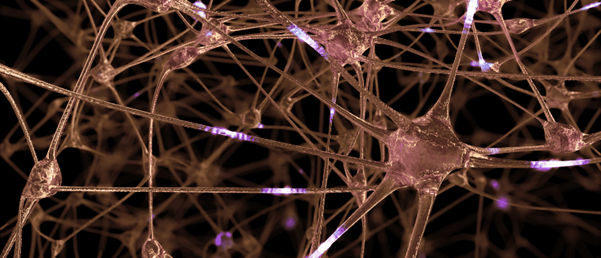
The researchers confirmed, through immunohistochemistry, that their tissue engineering method was able to generate dense three-dimensional axon networks that form connections between different cortical layers, which replicates the neural circuits in the brain. They also confirmed the presence of both spontaneous nerve-like activity and electrical- and pharmacological-evoked responses using calcium imaging and electrophysiological recordings.
Forsythe commented, “Not only were we able to construct a basic layout similar to what we see in regions of the brain, we found that the neurons actually behaved and performed in a similar manner.”