Knocking on nature’s door: bioinspired hemostatic agents
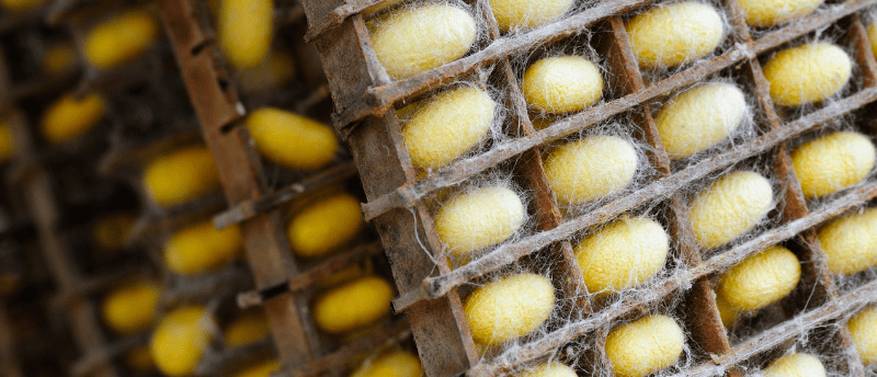
Proteins found in silkworm cocoons and mussels have inspired a novel hemostatic wound agent, which has proven successful in counteracting bleeding in a preclinical model of liver damage.
A recent study by researchers from the Pohang University of Science and Technology (Pohang, South Korea), Ewha Womans University (Seoul, South Korea) and the Catholic University of Korea Seoul St. Mary’s Hospital (Seoul, South Korea) has unveiled a novel material used in wound healing. The material, formed of a protein derived from mussels and silkworms, displayed effective tissue adhesion and hemostatic properties when tested in a rat model of liver damage. This could overcome the problems associated with conventional wound healing materials, like inadequate tissue adhesion and infection risk, thereby improving clinical outcomes.
One of the first phases of wound healing is hemostasis, which is the cessation of bleeding through a variety of mechanisms like vascular constriction, platelet aggregation and fibrin formation [2].
Hemostatic agents, like gauze or bandages, aid the process of hemostasis and reduce tissue damage. However, these commercial hemostatic agents suffer from several drawbacks, including inadequate tissue adhesion and insufficient prevention of contamination. Additional complications like inflammation and infection can arise if these materials become unintentionally embedded in the wound site.
Although these shortfalls can be prevented with the use of biodegradable substances, like fibrin glue and collagen sponges, these require proteins sourced from humans or animals, which inflate the costs associated with the medical procedures.
To develop hemostatic materials that overcame these challenges, researchers turned to proteins produced by mussels and silkworms, which have received considerable interest in the academic community because of their associated biocompatibility and efficiency in clotting blood.
The researchers developed a dual-sided nanofibrous wound dressing through electrospinning. The wound-adhesive inner layer was formed from mussel-derived adhesive protein and dihydroxyphenylalanine – a compound that promotes blood clotting. The anti-adhesive and hydrophobic outer layer was formed from methanol-treated silk protein derived from silkworms.
In vivo investigations using a rat model of liver damage demonstrated that the material reduced blood loss and clotting time while also being biocompatible with the tissue and biodegradable.
“We have validated the exceptional hemostatic performance of a multifunctional topical adhesive hemostatic agent that is derived from nature and is based on degradable proteins in the human body,” commented Hyung Joon Cha, co-lead of the study. “We will continue further research to assess its applicability in real-world patient care or surgical settings.”
While this study identifies a novel, dual-sided, hemostatic and protective material that could improve the medical tools available for wound healing, it also highlights that innovative materials can be found in nature.
You may also like:
 Finding a promising hydrogel glue in crustacean shells?
Scientists have utilized a polymer found in crustacean shells to improve the medical potential of hydrogels.