Load ‘em up: using nanoparticles to speed up diabetic wound repair

Researchers have designed a therapy to accelerate diabetic wound healing.
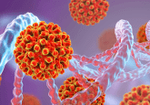
In a recent study, a team of researchers at the Icahn School of Medicine at Mount Sinai (NY, USA) has designed a trisulfide-derived lipid nanoparticle (TS LNP)-mRNA therapy to accelerate diabetic wound repair. The study detailed how the treatment targeted specific immune cells, reduced inflammation and reduced harmful molecules in damaged skin in mouse models.
Diabetic wounds are notoriously difficult to manage with current treatments due to persistent inflammation caused by dysfunctional macrophages and the unregulated accumulation of reactive oxygen species (ROS). When ROS are produced in excess, oxidative stress can occur, resulting in cellular, protein and DNA damage as well as inflammation. In the case of many individuals that face chronic inflammation from their diabetic wounds, macrophages that are supposed to initiate, maintain and resolve inflammation can be troublesome when they become dysfunctional. Inflammation that stems from dysfunctional macrophages not only damages surrounding cells but also hinders the wound healing process.
The team set out to address the critical health risk posed by diabetic wounds, which affect millions worldwide. Corresponding author Yizhou Dong explained the intention behind the study:
“Dysfunctional macrophages drive diabetic non-healing wounds, but we can reprogram them to stop the damage and instead help the healing process. We aim to promote faster and more effective wound closure by reprogramming these cells and modulating the inflammatory environment.”

Disc comfort: gene therapy for chronic back pain
An extracellular vesicle-based gene therapy has repaired damaged intervertebral discs in a mouse model of chronic back pain.
The team synthesized and formulated 98 different types of LNPs designed to respond to ROS. TS LNPs were loaded with mRNA encoded with IL4, a cytokine which plays a crucial role in regulating the immune response. These loaded TS LNPs, known as TS2-IL LNP-mRNA served two functions, primarily reducing the excess ROS at the wound site, helping to mitigate oxidative stress and tissue damage. Macrophages are then also able to take up the delivered TS2-IL LNP-mRNA at the wound site, leading to the production of IL4 protein within the macrophages. This IL4 prompts the macrophages to transition from pro-inflammatory M1 state to an anti-inflammatory M2 state, which is associated with tissue repair and wound healing.
The team tested this formulation in a diabetic mouse model, db/db mice. The treatment significantly improved wound healing, demonstrated by better epidermis formation, increased blood vessel formation and the presence of myofibroblasts.
The study’s findings indicate significant potential for treating diabetic, acute and chronic wounds. The TS LNP-mRNA platform provides an effective and beneficial therapeutic option. The team plans to validate its safety and efficacy in humans with a randomized controlled clinical trial.
“Our ultimate goal is to translate these findings into tangible benefits for diabetic patients. With further research and validation, this RNA-LNP therapy could potentially revolutionize diabetic wound management with one easily scalable application of a comparatively inexpensive therapeutic agent,” commented Dr. Dong. “The study also suggests the potential for RNA-LNP therapeutics to be more as pro-inflammatory macrophages are implicated in a wide range of diseases.”