Oxford University (Oxford, UK) researchers print 3D models of joints from bone specimens spanning 350 million years to better understand orthopedic problems

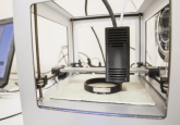
A team from Oxford University (Oxford, UK) has scanned over 200 specimens to produce CT scans which were used to print interactive 3D models of human joints. Researchers believe these models will allow better understanding of orthopedic complaints. Researchers from Oxford University (Oxford, UK) have created interactive 3D models of human joints in order to demonstrate how common medical issues may have occurred and how we are likely to evolve in the future. Using 128 slice CT scans of bones from humans, early hominids, primates and dinosaurs, the 3D computer models were compiled. A total of 224 bone specimens were...