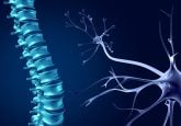
Reconnecting spinal circuits: neural progenitor cell transplantation shows promise for spinal cord injury
Neural progenitor cells with the ability to establish motor circuit connections in spinal cord injuries have been identified.
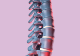
A recent study, conducted by researchers at Texas A&M University (TX, USA), has identified a rare group of neurons that are capable of reconnecting broken spinal circuits and activating leg movement in mouse models of spinal cord injury. Could these identified cells help enhance stem cell therapies for paralysis following spinal cord injuries?
For years, scientists have hoped to transplant neural stem cells into injured spinal cords to rebuild lost connections. However, identifying the precise cells within these grafts that can integrate with the spinal cord’s walking circuits has been a significant challenge. In the United States alone, hundreds of thousands of people live with spinal cord injuries and there are currently no FDA-approved therapies that can restore neurological function. This new study may bring scientists closer to improving stem cell therapies by identifying effective neural stem cells capable of reconstructing damaged spinal motor circuit pathways.
Senior author Jenifer Dulin explained the team’s approach: “What we’re trying to do is place new cells into the middle [of a disconnected spinal motor circuit] so they can reconnect the pathway and allow signals to flow again.”
 Advanced organoids reveal new pathways for spinal cord repair
Researchers have created the most advanced human spinal cord organoid model to date.
The researchers transplanted neural progenitor cells into the injured spinal cords of mice and studied how these cells formed connections to surrounding nerve networks. Specifically, they examined how graft-derived neurons integrated with spinal motor circuits responsible for controlling hind limb (back leg) movement.
The results were promising. When researchers experimentally activated a select group of transplanted neurons, the hind limb muscles responded, indicating that the grafted cells had effectively integrated into the motor circuitry of the spinal cord. However, the team soon realized that these newly identified interneurons were quite rare within the transplanted cell population. In approximately 20–30% of the animal models, leg muscle responses were observed, but it was enough to be meaningful data.
“This is meaningful because it shows the potential to re-create these walking neural circuits is there,” Dulin explained.
The findings could play a pivotal role in paving the way for the next generation of regenerative therapies by identifying specific neurons that need to be enriched in transplanted cell populations. Additionally, the research highlights the importance of rehabilitation as a factor that may influence recovery outcomes.
Dulin explained that newly transplanted neurons require time to adapt to the spinal cord’s environment, requiring activity.
“We’re essentially putting newborn neurons into the spinal cord, and they don’t have any experience yet,” elaborated Dulin. “… these transplanted neurons need activity to learn how to function within the circuit.”
Looking ahead, the team aims to further explore their discovery to uncover why certain animals responded to the treatment while others did not.
“This kind of basic biology research is critically needed in order to develop new therapies,” Dulin said. “For decades in the field of spinal cord injury we’ve just been testing treatments without really understanding how they work. We’re entering a new era where we have amazing tools to really study the effects of a treatment on an individual cellular level. These kinds of studies are critical to paving the way for effective human treatments.”