Seeking the genetic instruction manual for limb regeneration & wound healing

In this interview, Drs Benjamin L King (Senior Staff Scientist) and Voot P Yin (Assistant Professor) from MDI Biological Laboratory in Bar Harbor (ME, USA), discuss their discovery of a common miRNA-regulated genetic network of genes controlling limb regeneration in regenerative species and the path ahead, as well as the implications for this research and some of the wider roles miRNAs play in regeneration.
Extensive regeneration occurs in certain animals such as amphibians and teleosts, allowing them to fully regenerate body parts such as their limbs, tails and retina. Despite humans sharing many of these genes within these organisms, our adult regenerative capacity is limited.
Drs Benjamin L King and Voot P Yin at the MDI Biological Laboratory in Bar Harbor (ME, USA) carried out a next-generation sequencing analysis into the genetics of these organisms to identify the genetics regulating blastema formation — the key step in regeneration in animals such as the zebrafish, axolotl and the bichir, where injury the injury site forms a mass of dedifferentiated cells that proliferate and differentiate in a coordinated manner to re-form the lost appendage.
Their analysis of their RNA-seq transcriptome demonstrated an evolutionary divergence approximately 420 million years ago of a common miRNA-regulated genetic network of blastema genes, identifying new genes involved in limb regeneration and a significant role of miRNAs, suggesting that regeneration is an ancestral trait that could be reactivated in humans.
In this interview, Drs Benjamin L King and Voot P Yin from MDI Biological Laboratory in Bar Harbor (ME, USA), discuss their discovery of a common miRNA-regulated genetic network of genes controlling limb regeneration in regenerative species, the implications and potential applications of this research, the wider role of miRNAs in regenerative processes, and the road ahead.

Voot P Yin
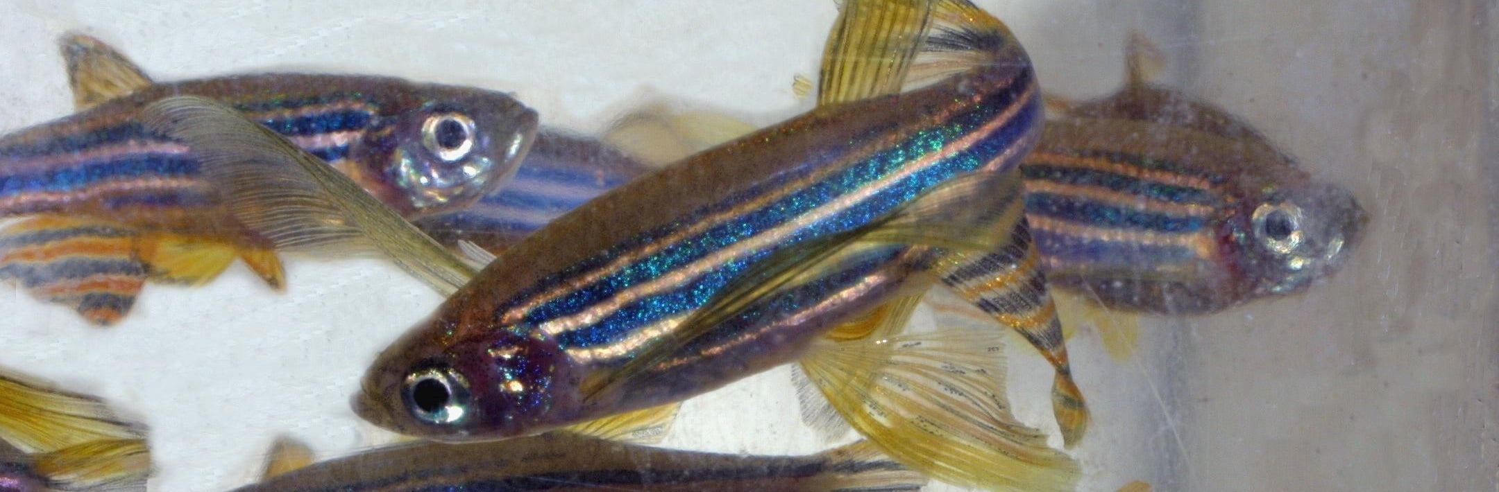
Dr Yin is studying limb and heart muscle regeneration in adult zebrafish. Humans share the same genetic program to regenerate missing or damaged tissue as zebrafish, but the genetic code has been deactivated. He is seeking to understand how zebrafish regenerate damaged tissue so that therapies can be developed to reawaken the dormant genetic codes for regeneration in humans. Using the adult zebrafish, Dr Yin has discovered that miRNAs play a central role in the initiation and propagation of this process. Furthermore, his lab has recently discovered that MSI-1436, a repurposed drug, enhances tissue regeneration of complex limb and heart tissues. This groundbreaking work led to the co-founding of Novo Biosciences, Inc. (Bar Harbor, ME, USA), a regenerative medicine company devoted to accelerating our innate ability to heal. Dr Yin currently serves as Novo’s chief scientific officer. His research has implications for limb regeneration, wound healing, prosthetics, heart muscle regeneration and muscular dystrophy.
Email: [email protected]
Telephone: +1 207 288 9880 ext. 474
Benjamin L King
Recent advances in genomics are transforming how we study biology, making it essential to gather and analyze increasingly large volumes of data. Dr King specializes in bioinformatics, in which data are analyzed and integrated with existing knowledge. He applies this data-intensive discovery approach to the study of the fundamental mechanisms of regeneration. The ability to mine unexplored data promises to speed the pace of discovery in the biomedical sciences. His team is developing the Comparative Models of Regeneration Database (RegenDB), which provides a systems-level view of tissue regeneration models to advance knowledge of regenerative biology and stem cell self-renewal. Scientists can use RegenDB to analyze integrated functional genomic datasets of regenerative processes to identify conserved gene networks within and across species. Under Dr King’s leadership, the MDI Biological Laboratory is seeking to become a center for education in data-intensive discovery.
Email: [email protected]
Telephone: +1 207 288 9880 ext. 139
Can you tell us about your careers to date and how you came to be involved in researching the genetics of appendage regeneration?
Ben King: My background is in genomics and bioinformatics — a particular discipline that is broadly utilized across medical sciences as well as biology in general. We now have the ability to characterize the complete genome and gene activity of essentially any organism: it’s an incredibly powerful approach and does require some specialized expertise, which I have acquired over the years.
My undergraduate and masters degrees were in biomedical engineering from Boston University (MA, USA) and my department later launched one of the first graduate programs in bioinformatics. I initially worked on protein structure modeling and computer-aided drug design, but my career followed the revolution in genomics.
The opportunity to study genomes of non-model organisms for the first time, especially in the context of regeneration, led me to MDI Biological Laboratory 7 years ago. High-throughput sequencing has enabled us to study animals that have an inherent ability to regenerate virtually any tissue and learn essentially how those mechanisms work, and then of course compare to mammalian tissues, which typically have less regenerative capability.
Voot Yin: I started my scientific career at the University of Utah in Salt Lake City (UT, USA) where I earned a PhD in the department of Human Genetics. I focused on understanding the process of tissue destruction and remodeling during normal animal development, and how key genetic players regulate these processes. So at a very early stage in my career I recognized the fact that animals have really perfected cellular processes that we’re interested in today.
From then I went to Duke University Medical Center (NC, USA) where I joined the lab that pioneered zebrafish as a model system to interrogate both appendage and heart muscle regeneration — that’s what I do now in the lab. My interest in microRNAs (miRNAs) spans from the desire to define the genetic circuits that allow regenerative processes to occur very well in organisms such as the zebrafish, axolotl and the bichir. These animals have the innate ability to regenerate limbs and the genetic underpinnings have been subjected to evolutionary pressures for hundreds of millions of years. We would like to identify these critical genes and manipulate them in humans in order to improve our own regenerative capacity.
The focus with appendage regeneration was to decode and to reduce the number of potential genetic influences down to a very small number. Given that miRNAs are conserved at the sequence level across all organisms it should be easy to compare and identify common factors.
Can you summarize the role you found miRNAs to play in the initiation and propagation of the regenerative cascade of limbs in your recent study?
BK: miRNAs are highly conserved and known to be post-transcriptional master regulators of many other genes, so we wanted to look at the miRNAs in the regenerative responses in the regenerative organisms, zebrafish, axolotl and the bichir. We chose these organisms because they regenerate appendages in the same way at a cellular level. After clotting and wound healing following an injury in these animals a tissue, called the blastema, forms over the wound. The blastema cells are highly proliferative and the blastema is maintained throughout outgrowth in all three organisms; furthermore, in different model systems if you inhibit the formation of the blastema, regeneration cannot occur.
We therefore wanted to look at that equivalent critical time point of blastema formation in all three systems. We found that some of these miRNA were highly upregulated and five of them were downregulated, and these miRNAs can of course mediate a certain battery of genes and regulate them. So, we have a model where the miRNAs circumvent the top of the process and control the networks of protein-coding genes that are required for blastema formation.
VY: A key principle that we follow in developmental biology in these types of profiling studies is that an injury to an animal system is reflected at the genetic level. Therefore, genes that are either significantly upregulated or downregulated during tissue injury probably have an important role in mediating the repair process. That is why for us, the focus has been on the miRNA that are significantly upregulated in the context of blastema formation.
“miRNAs are highly conserved and known to be
post-transcriptional master regulators of many other genes, so we wanted
to look at the miRNAs in the regenerative responses in the regenerative
organisms, zebrafish, axolotl and the bichir.”
What were the biggest challenges and the biggest surprises in the study?
VY: When you look at the position of these three animal systems in the evolutionary tree they last shared a common ancestor about 420 million years ago. Each appendage has different tissue composition and rates of regeneration, yet when you look at the critical structure, the blastema, there’s actually a well-conserved set of miRNAs. To me it is surprising but also revealing, as it tells us that if we want to manipulate or enhance blastema formation in a non-regenerative system we now have the key ingredients that have been favored during evolution as being critically important.
BK: With the high-throughput sequencing we could really understand for the first time what miRNAs and genes are expressed in all three systems. The zebrafish of course is a widely utilized model organism with a well-characterized genome and many studies have been carried out looking at caudal fin regeneration in the zebrafish, but with the axolotl there are fewer genetic tools and its genome is estimated to be 10-times as large as the human genome. Then another challenge is of course working with Polypterus senegalus (bichir) as there was essentially no previous knowledge to rely on, so a challenge was trying to assemble what we call a transcriptome and identify miRNAs in both the axolotl and the bichir.
In particular, seeing miR-21 consistently upregulated and the most highly expressed miRNA in all three systems was one of the biggest surprises and exciting to see. It demonstrates just how powerful genomics is by characterizing organisms with traits of interest rather than confining studies to traditional model organisms.
Have you found miRNAs to play a significant role in regeneration of other organs aside from limbs or appendages?
BK: Yes for sure. Voot has done a lot of work over the years with both appendage and heart regeneration — the first miRNA he found to be required for regeneration was an miRNA called miR-133 back in 2008 for caudal fin regeneration in zebrafish and of course blastema formation. What is striking is he also found miR-133 to be required for heart regeneration in zebrafish. He recently published a study in Development demonstrating that miR-101 is also required for zebrafish heart regeneration.
VY: When one looks at miRNAs, what has been surprising is that by and large the individual miRNAs are dispensable for normal development, but then when you have an injury or trauma situation in adult tissues the role of these miRNAs are revealed. We are finding out that miRNAs like let-7, miR-21, miR-101 and miR-133 actually have important roles in a number of different tissues during the repair and regeneration process.
Ben spoke about the appendage and heart — other groups have shown important roles of miRNAs in lens, spinal cord and liver regeneration. I think miRNAs are really genetic readouts of tissue damage and their activation is the first domino in tissue repair and regeneration, so it’s not surprising that a single miRNA can have multiple roles in various tissues.
“When one looks at miRNAs, what has been surprising is that by and large the individual miRNAs are dispensable for normal development, but then when you have an injury or trauma situation in adult tissues the role of these miRNAs are revealed.”
Some mammals such as mice have increased regenerative capacity as neonates compared with as adults. Do we know why this is, and if the mechanism relates to the one you identified in your study?
VY: I think the best example that speaks to this is actually not appendage regeneration but heart regeneration. By recapitulating the work performed in adult zebrafish, a number of years ago Eric Olson’s group at University of Texas Southwestern (TX, USA) showed that 1-day-old mice had this remarkable ability to regenerate heart muscle, but a week after birth that ability is lost because of changes in gene expression. Many of these genes were actually miRNAs.
There are a lot of ideas as to why regenerative capacity goes down during development and during aging. One is you’re getting maturation and changes in tissues and organs that are needed in order to support the larger workload that an adult organism requires. The other common idea is that the development and maturation of the immune system is a critical competent, as these processes coincide with the decrease in regenerative capacity. I’m sure miRNAs have played a pivotal role in both of these processes.
BK: Yes, I think there is a lot of work that continues to be done to study neonatal mouse heart and its regenerative capacity. Learning what mechanisms turn off that regenerative capacity is important.
“There are a lot of ideas as to why
regenerative capacity goes down during development and during aging. One
is you’re getting maturation and changes in tissues and organs that are
needed in order to support the larger workload that an adult organism
requires. The other common idea is that the development and maturation
of the immune system is a critical competent.”
As the genes identified in your research are present in humans, what are the steps to find out if this regenerative mechanism could be activated in humans?
BK: As you stated, all of these genes are present in the human genome, but they are regulated differently, so finding mechanisms or therapies that could reactivate them is critical.
VY: A lot of biology is dictated by the timing and location of the activation of genes. For example, we have an idea of the changes in overall levels of certain miRNAs and for some of them we often know when and where they are turned on, at least in highly regenerative systems, but we don’t yet know what the situation is in non-regenerative systems like humans.
We know that regeneration is an orchestra of communication signals from a number of different tissues, and those tissues have to turn on genetic light switches at the same time, so part of the challenge in defining those circuits in greater detail is going to be interrogating these miRNAs and the potential target genes in a mammalian system. Is the order and location of gene activation similar? If not, then we have clues as to what we need to manipulate in order to reactive these circuits.
“We know that regeneration is an orchestra of communication signals from a number of different tissues, and those tissues have to turn on genetic light switches at the same time, so part of the challenge in defining those circuits in greater detail is going to be interrogating these miRNAs and the potential target genes in a mammalian system.”
What do you think a realistic timeline for this is?
VY: This is a tough question because it is very much like peeling apart the layers of an onion: you never known how many more layers you need to peel in order to get to the core. We are also limited by the complexity of biology and research funding.
Biology is incredibly complex system and as soon as we open one door there are 500 million more doors that we need to at the very least tap on. So I think the possibility of enhancing regeneration or limiting tissue degeneration is probably within 5—10 years. The reason why that is important is then that opens up the door for interfacing with other technologies that exist today — combining prosthetics and tissue engineering as a way to enhance the interface between an injured or amputated limb with a synthetic technology and prosthesis.
BK: It is hard to think about a timeline for being able to activate these regenerative mechanisms in humans as even if we had a small-molecule drug that worked in a mouse it would take a very long time to get that through clinical trials and out as a product. We can learn a lot by trying to inform the development of those therapies of course and by comparing these systems using a similar approach but in organisms that don’t regenerate as well.
If you look at injuries after ischemia in the heart in a zebrafish and human, the initial events are similar but the zebrafish heart can resolve the scar tissue and restore heart function. If we had some way of resolving scar tissue in humans, even by some fraction, it would be a huge advance. It wouldn’t be equivalent to regenerating an entire new heart but it would have great benefit for millions of individuals with heart disease.
“It is hard to think about a timeline for being able to activate these regenerative mechanisms in humans as even if we had a small-molecule drug that worked in a mouse it would take a very long time to get that through clinical trials and out as a product.”
How do you hope to translate this research to wound healing?
BK: In our studies we have looked at time points before blastema formation, so we have data on the miRNAs being upregulated and downregulated when the wound epithelium is formed during wound healing. So, the overall approach of profiling miRNA and other genes could be used to find a conserved regulatory circuit for other stages of regeneration including wound healing.
VY: I think what we are doing in terms of studying limb regeneration and blastema formation in these highly regenerative systems is highly relevant to wound healing, because blastema formation is effectively wound healing in hyper-overdrive.
What I mean by this is that wound healing occurs in all organisms: what we are finding out with salamander, Polypterus and zebrafish limb regeneration is that the process of wound healing is so rapid that it actually minimizes or suppresses scar formation. In humans the dominant healing mechanism during wound healing is the formation of scar tissue. The scar can actually be detrimental in the long run, so by studying processes of limb regeneration and blastema formation I think we will identify potential ways in which we can speed up wound healing and actually minimize the potential of scar tissue formation in humans.
You mentioned that a more near-term application for this knowledge could be the ability to regenerate some of the nerves at an amputation point, allowing prostheses that interface with the nerves for improved control. Are there any other potential applications you foresee at this time?
VY: I think when you’re talking about an amputation there is a fair amount of pain, so one thing you can imagine is that if the process of wound healing and regeneration is enhanced so that time period of pain would be reduced, as well as reducing the risk of infection. With any type of surgical procedure, surgeons will tell you the most common thing that results from that is abnormal healing caused by secondary infection or elevated levels of scar tissue formation. All of these things could be minimized if not completely eliminated, if the rate of wound healing and the enhancement of regeneration is stimulated in patients. This results in improving the patient’s quality of life and effectively reducing the healthcare expenses that result from initial injury.
BK: As we find mechanisms that control key events that are necessary for regeneration, these can be translated to many other aspects of biology. Cellular proliferation for example is important during outgrowth and other events during regeneration, and being able to control cellular proliferation is relevant to cancer. There are many fields that can benefit from our studies of regeneration, but of course we can benefit from what other groups study — whether cancer or other aspects of biology.
“As we find mechanisms that control key events that are necessary for regeneration, these can be translated to many other aspects of biology.”
Do you have any idea of how a potential therapy for appendage regeneration might look, for example as a small-molecule therapy, gene therapy or a combination of different approaches?
VY: For tissue regeneration I think the approach I favor is small molecules as they can be incredibly specific for the target gene and we can actually make cocktails of small molecules to target an entire pathway or multiple target genes from different pathways that might be critical for regeneration process.
I think one of the most understated benefits of small molecules is that you can dial in the efficacy. In other words you can increase and lower the dose, and if any of these compounds have adverse effects you can just stop administration and the problem goes away. In contrast, stem cell biology and gene therapy are more permanent solutions, and as we’ve seen in the last 20 years of regenerative therapy approaches that have been focused on stem cells and gene therapy, there are problems that we have yet to overcome.
The idea of small molecules is wonderful because you are now tapping in to the natural instruction manual that exists in all of us: the natural DNA program that resides in all of our cells and we would just simply reactivate these endogenous programs and not reinvent the wheel.
Voot, you and colleagues Kevin Strange and Michael Zasloff have just been awarded a patent for use of the small molecule MSI-1436 to stimulate the repair and regeneration of heart tissue damaged by injuries such as a heart attack. What is the significance of this for you?
VY: Our patent for MSI-1436 is great validation for my lab’s approach to improve regenerative capacity in humans. We initiated these studies with the zebrafish and then applied our findings into the adult mouse. Normally injuries to the adult mouse triggers scar formation and a gradual decline in heart function: MSI-1436 enhanced heart regeneration in the zebrafish and also improves heart function in the adult mouse. Theses findings show that the regenerative circuits that the zebrafish employs has been conserved throughout evolution. If MSI-1436 works in humans, it will be a game-changer for patients with heart disease.
What do you both hope to have achieved in the next 5—10 years in your careers?
BK: To have studied a very broad sampling of different animals that have a range of regenerative capacities, so that we can better understand why some animals are inherently better at regenerating tissues than others, would be a fantastic achievement. It’s something that we’re just scratching the surface of with our study.
VY: I’ve really been interested in decoding the genetic blueprint to stimulating tissue regeneration in both limbs and the heart. I would love to expand upon this by identifying a handful of critical target genes and then small molecules that can augment the expression and activity of those genes. That will allow us to take the lessons we have learned from these lower vertebrates and apply them to see if we can enhance regenerative capacity in humans — I think that’s what we are all after. I do think it is possible with the combination of developmental biology and the technology advances in bioinformatics that Ben spoke of.
Finally, if you had unlimited resources to carry out any work, what would it be?
BK: I would not only study miRNAs and their role in regeneration but more broadly look at other non-coding RNAs (ncRNAs). We’ve gathered a lot of data on miRNAs, but there are longer transcripts generated from non-coding genes that also have the potential to regulate other genes. It is really unchartered territory as to how these longer ncRNAs work, so it is important to interrogate the function of this larger class of RNAs. In short, I would aim to fully understanding the role of ncRNAs and the epigenome of regeneration.
VY: If I had unlimited resources to carry our any work, I would do two particular studies. First, I want to assemble an interdisciplinary team including developmental biologists like myself who rely on animal systems to provide important cues of regenerative capacity, with members of the team who are focused on tissue engineering. I’d also have a group that looks at engineering from a mechanical perspective and I want to merge all of those disciplines because I believe that with a topic like appendage regeneration you need the expertise from all of these different groups to really push the frontier of limb regeneration.
However, as an intermediate step before we are fully able to fully regenerate limbs there is the unique opportunity to bring in engineering to create systems that people often refer to as artificial intelligence, prosthetics — I’d love to get that as a big umbrella of regenerative medicine to help improve both the function of limbs as well as the quality of life in patients.
Second, I’ve really been interested in the idea of energy usage during states of regeneration. To be able to replace any damaged tissue to the original function and form is obviously pretty darn useful to have, so why has it been modified during development and evolution in humans? One of the ideas is that there is an energy benefit/cost that comes with this ability. I want to understand this potential tradeoff a little bit more. I want to understand how animals utilize energy in an uninjured state and how this may shift in the injured environment, and whether that type of change in energy usage is also evident in humans.
Do you have any other comment’s you’d like to add?
BK: The paradigm of data-driven discovery is something that I think will grow over time and will continue to have a leading role in here at MDI Biological Laboratory. Essentially all labs in the world are applying some aspects of genomics to their analyses, and having more people trained in using these tools is important.
VY: I’d like to go back to a point that I hinted at earlier — the idea that evolution has really created a beautiful canvas for understanding regeneration with all these different organisms that already exist in nature, and that we could learn quite a bit of information by querying not only individual systems but multiple systems that share this unique quality in biology. By doing that we are refining the important genetic factors to allow these processes to happen — this is something that we in the developmental and scientific community refer to as comparative biology — comparing different systems to understand an aspect of biology.
This is something that is reemerging in the context of scientific research but also something that we do very well at this institute and it is the backbone and foundation of the MDI Biological Laboratory. We are building upon that foundation and bringing in the state of the art technology in bioinformatics. It is this combination that is enabling these major advances in regenerative biology and medicine and I think that will be a common theme moving forward.
“The paradigm of data-driven discovery is something that I think will grow over time and will continue to have a leading role in here at MDI Biological Laboratory.”
Further reading
- King BL, Yin VP. A conserved microRNA regulatory circuit is differentially controlled during limb/appendage regeneration. PLoS ONE doi:10.1371/journal.pone.0157106 (2016).
- Beauchemin M, Smith A, Yin VP. Dynamic microRNA-101a and Fosab expression controls zebrafish heart regeneration. Development 142(23), 4026—4037 (2015).
- Yin V, Smith A, Roberts H, Carlisle H. ZF143 enhances zebrafish heart regeneration and improves mouse heart function after an LAD injury. FASEB J. 29(1) Supplement 1029.10 (2015).
- https://mdibl.org/press-release/from-sci-fi-to-reality-unlocking-the-secret-to-growing-new-limbs/
- https://mdibl.org/press-release/mdi-biological-laboratory-scientists-awarded-patent-for-potential-new-heart-disease-drug/
Images via MDI Biological Laboratory in Bar Harbor (ME, USA).