Advancing multiple sclerosis with cerebral organoids: an interview with Nicolas Daviaud

With this year marking ten years since the development of cerebral organoids, we spoke to Nicolas Daviaud to learn how the Tisch Multiple Sclerosis Research Center of New York (USA) is using them as a model for multiple sclerosis (MS).
Dr Daviaud also explains the unique features of cerebral organoids that make them particularly useful for investigating the factors that might trigger or facilitate disease onset, such as the much-discussed link between Epstein-Barr virus and MS.
Can you provide an overview of your research with cerebral organoids to study and develop treatments for MS?
We are developing cerebral organoids from patients’ cells. Therefore, our organoid contains the genetic information of the patients they were derived from. Cerebral organoids recapitulate early human neurodevelopment, including the generation, proliferation, and differentiation of neural progenitors into glial cells and neurons and their interactions. Thus, this innovative model of MS allowed us to study the patient’s genetic contribution to MS onset and development, but also its interplay with environmental triggers, such as viral infection. This work will hopefully lead to the discovery of new mechanisms and potential therapeutic targets.
What advantages do organoid models offer over alternative methods in understanding the underlying mechanisms and potential therapeutic targets for MS?
Organoids offer a more complex and more accurate representation of the human brain compared to regular two-dimensional cell cultures. Firstly, organoids contain multiple organ-specific cell types. In our case, these include neural stem cells, neurons, astrocytes and oligodendrocytes. Secondly, all of those cells are grouped together and spatially organized in a similar manner to the human brain. Finally, organoids are capable of recapitulating some specific functions of the organ, such as neuron activity. This makes cerebral organoids a very useful tool to study neurological disorders, such as MS.
Animal models have been enormously helpful over the past several decades in our overall understanding of MS. However, mouse models cannot replicate the complexity of the human brain. Moreover, MS is a human disorder, and mice cannot naturally develop the disease, making the existing rodent model inaccurate. It is also important to note that cerebral organoids do not contain immune cells, and thus do not reproduce the inflammatory demyelination classically associated with MS. We used this as an advantage to understand the genetic variant liability in MS pathogenesis[1]. The use of cerebral organoids also allows for the control of the microenvironment in which the brain develops. We can then choose to add or remove factors that might be associated with MS, such as exposure to Epstein-Barr virus, vitamin D, or even nicotine.
For these reasons, the use of a human cerebral organoid model of MS might lead to new discoveries regarding MS development in patients.
What are some of the key findings that have been gained through your research using cerebral organoids in the context of MS?
We describe cerebral organoids as an innovative model to study MS and how the patient’s genetic background leads to differences in brain development that might trigger or facilitate the disease onset.
We detected a decrease in proliferative capacity, notably in progressive forms of MS, associated with a reduction in the progenitor pool and an increase of neurogenesis possibly due to an asymmetric shift of the cell division mode. We also observed a decrease in glial cell development, particularly oligodendrocytes (the cells that make myelin). This dysregulation seems to be due to a defect in the progenitor’s cell cycle. We linked these effects to a strong decrease of p21 expression in Primary Progressive MS organoids, unrelated to DNA damage or the apoptosis pathway.
With 2023 marking ten years since the development of the first cerebral organoids, what challenges need to be overcome and where do you see this field heading in the next decade?
Despite the advantages of using organoids to study disorders, some challenges remain. Variability in organoid development is a known confounding factor. However, new, improved protocols are being developed to overcome this limitation. The lack of blood vessels and immune cells also prevents organoids from providing a complete overview of brain development. However, new protocols are also being tested to integrate these features into cerebral organoids. It is important to note that organoids remain an in vitro model and they do not interact with other organs. Therefore, they are not able to replace in vivo models.
In just one decade, the number of research articles using organoids has increased significantly. Protocols have been developed to grow organoids of almost every organ. We can now create a beating heart, lung, kidney, brain, or even gut organoid. In a recent press release, the Tokyo Medical and Dental University (Japan) announced the first successful transplantation of an organoid into a patient with ulcerative colitis.
Cerebral organoids from different regions of the brain can also be created and “fused” together to create an even more complex model of the human brain. Recently, Madeline Lancaster’s (Cambridge University, UK) group showed that organoids have the capacity to exhibit extracortical projecting tracts that can innervate a dissected mouse spinal cord and trigger the contraction of a muscle.
In just ten years, organoids have shown incredible potential to create innovative human models. They can provide new insights into the onset and development of disorders, and they may also be used as a new therapeutic approach through personalized tissue transplantation. It is very difficult to predict what will happen in the next ten years, but organoids are sure to play an integral role in biomedical research.
Meet the interviewee
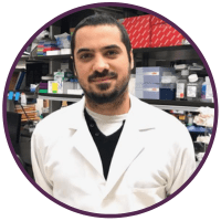
 Nicolas Daviaud, Research Scientist, Tisch MS Research Center of New York (USA)
Nicolas Daviaud obtained his PhD in neuroscience from the University of Angers (France) in 2013 prior to joining the Icahn School of Medicine at Mount Sinai (NY, USA) as a postdoctoral researcher where he worked with cerebral organoids to model preterm hypoxic injury and performed cerebral organoid transplantation to repair brain lesions. In 2019, Dr Daviaud joined the Tisch Multiple Sclerosis Research Center of New York (USA) as a Research Scientist. Here, he leads a research project on the use of cerebral organoids to study MS.
Disclaimer
The opinions expressed in this interview are those of the interviewee and do not necessarily reflect the views of RegMedNet or Future Science Group.

