GMP-compliant iPSCs show clinical potential

Induced pluripotent stem cells manufactured using a novel, current good manufacturing practice (cGMP)-compliant, non-lineage specific process could have clinical potential.
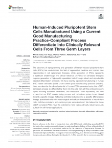
Research into human induced pluripotent stem cells (iPSCs) has demonstrated they can be differentiated into all three embryonic germ layers, the ectoderm, endoderm and mesoderm. The current good manufacturing practice (cGMP)-compliant directed differentiation was unbiased in terms of specific lineages, and was found to be robust and reproducible.
iPSCs have shown huge potential for drug and disease modelling as well as the production of therapeutics, but consistency and reproducibility have remained challenging. In particular, the robust requirements of cGMP have proved costly and complex to achieve.
In the paper, the authors commented “There are still major obstacles toward clinical application of human iPSC: high cost of production and post-transplantation immunological reaction, which highlights the need for generation of large HLA-matched MCBs from donors with selected haplotypes.”
In this work, human iPSC lines were generated using cord blood-derived cells following morphological and biomarker assessment. They differentiated into ectoderm, mesoderm, and endoderm through embryoid body (EB) formation in an unbiased manner, demonstrating clinical potential for allogeneic transplantation.
“We have shown that using optimized culture conditions it is possible to retain the ability of iPSCs to differentiate into three lineages when starting from the same cGMP-compliant starting material and that separate MCB banks may not be required. This can be a huge cost saving strategy in development of high-quality iPSC-derived products”, continued the authors.
Further research is requested to assess the influence of the epigenetic memory, donor cell source, state of pluripotency and number of reprogramming factors on the lineage differentiation and characteristics of terminally differentiated cells.
Click on the image below to download the pdf:
