How injuries are altered to regeneration signals at the molecular level
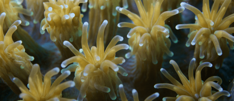
The process of regeneration was identified in the freshwater polyp, Hydra, over 200 years ago. However, it was previously unknown how the orderly regeneration of damaged tissues or organs is started following damage. An interdisciplinary research team at Heidelberg University (Germany) was able to demonstrate how wound healing signals emitted after damage are translated into precise signals of pattern formation and cell differentiation by studying Hydra. Mitogen-activated protein kinases (MAPK) and the Wnt signaling pathway are critical components that have remained remarkably unaltered throughout evolution.
Regenerating capabilities broadly vary in animals and most mammals and vertebrates have limited regeneration capacity. However, early-evolving basic and simple creatures, such as cnidarians and planarians, can renew their entire body. The genesis of the regeneration process starts with wound healing. Cells start to multiply and generate an undifferentiated mass – a blastema – from which the missing structures are re-patterned and formed. This triggers the same genetic pathways that regulate embryonic development.
To ascertain the implicated molecular pathways, the research team headed by Thomas Holstein examined the freshwater Hydra to grasp the fundamental principles of regeneration activation. The Hydra is split in half, causing the top half to regenerate a new ‘head’ and the bottom half to regenerate a new ‘foot’ – thus, completely distinct body parts can develop from the same tissue at the cut area in the center. The researchers at Heidelberg University’s Centre for Organismal Studies (COS) have now established how this is achieved, expanding on their previous work on Hydra regeneration.
Any type of damage, regardless of where it originates, generates nonspecific signals for an injury response, such as wound healing via calcium ions and the formation of reactive oxygen species. Three mitogen-activated protein kinases – p38, JNKs and ERK – transmit the signals intracellularly; both head and foot regeneration require the activation of these three molecules. Following this, Wnt signaling pathways are then activated, which are necessary for the creation of basic organs and the body axis during embryonic development. Thus, general wound healing signals are converted into position-specific patterning and cell differentiation signals for regeneration.
“Our experiments show that the Wnt signalling pathway is a main component of the initially general injury response and, depending on signal strength, directs the tissue toward head or foot development,” describes Holstein. This explains why, in the case of MAPK inhibition, artificially created, recombinant Wnt proteins can cause regeneration that would otherwise be missing.
“It was also surprising that in middle body parts that had both head and foot removed, heads can be induced at both ends in this way,” explains Suat Özbek, a representative of Holstein’s molecular evolution and genomics research group at the COS.
Wnt/-catenin, a component of the Wnt signaling pathway, was originally established to encode positioning information for the creation of new head structures. The research team of Holstein and Özbek collaborated with a team of mathematicians led by Anna Marciniak-Czochra to construct a model that explains how basal positional information in the tissue is converted through the Wnt signaling pathways from an initially undifferentiated damage response into a diverse patterning process.
“Because MAPKs and Wnts are highly evolutionarily conserved, this mechanism is likely deeply embedded in our genome, which is important for regenerative processes in vertebrates and mammals as well,” emphasizes Holstein.
The research article proposes that these described mechanisms are profoundly buried in our human DNA due to the high level of evolutionary conservation of MAPKs and Wnts.
Source: https://www.pnas.org/doi/full/10.1073/pnas.2204122119
Press release: https://www.sciencedaily.com/releases/2022/09/220902111335.htm