Hydrogel-based scaffolds: developing a strong support network for your brain
Researchers develop an electrically conductive hydrogel scaffold that is able to support neural cell differentiation ex vivo.
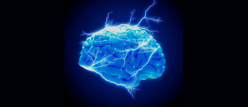
Brain-computer interfaces (BCIs) aim to link human brains to machines through electrodes implanted within the brain. BCIs could improve brain activity monitoring in patients with neurological conditions. However, due to their need to conduct electricity, BCIs are most often made of metal, which does not mimic the environment that brain cells typically grow.
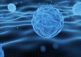
Now, researchers at the Wyss Institute at Harvard University, Harvard’s John A. Paulson School of Engineering and Applied Sciences (SEAS) and MIT (all MA, USA) have manufactured a type of electroconductive hydrogel scaffold, which resembles the soft, porous characteristics of brain tissue. They demonstrated that the scaffold promotes the proliferation and differentiation of human neural progenitor cells (NPCs) into a variety of brain cell types for up to 12 weeks.
In 2021, the research team manufactured a prototype hydrogel-based electrode, with the goal of creating a material that could bend and curve according to the brain’s structure. They showed that this electrode was compatible with brain tissue; however, to improve upon the design, the team decided to combine living brain cells into the electrode itself, as the most compatible material for live brain cells, are cells themselves. They thought this could allow electrical impulses to be transferred to a patient’s brain through more natural means of cell-cell interaction.
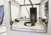
The researchers also added a freeze-drying step to the manufacturing process to create a porous scaffold, which led to improved cell proliferation. This is because the cells had a sufficient surface area on which to grow and allowed for the electroconductive components to form a continuous pathway through the hydrogel, to conduct nerve impulses.
The researchers created hydrogels that were either viscoelastic (like jelly) or elastic (like a rubber band) or soft or stiff. Once the scaffolds were created, they cultured NPCs onto the scaffolds to determine the physical properties that supported neural cell growth and development most effectively.
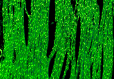
After 5 weeks, NPCs on the softer and viscoelastic hydrogel scaffolds differentiated into several different cell types with specific features, whilst those on elastic gels remained largely undifferentiated. Additionally, hydrogels that were made with larger amounts of conductive materials formed more branched networks.
To observe how electrical stimulation affected cell development, the researchers electrically stimulated them via the conductive materials within the hydrogel scaffold. Electrical pulses were supplied 15 minutes at a time, either daily or every other day. After 8 days, the scaffolds that had been stimulated every day had very few living cells, whilst those that had been pulsed every second day were densely packed with living cells.
The cells were left in the scaffold for 51 days following the 8-day period of electrical stimulation. The cells in the scaffolds that received daily electrical stimulation did not differentiate into other cell types, whilst cells that were stimulated every other day produced highly differentiated neurons and astrocytes with long protrusions. The difference in electrical impulses examined had no influence on the quantity of myelin produced.
“The successful differentiation of human NPCs into multiple types of brain cells within our scaffolds is confirmation that the conductive hydrogel provides them the right kind of environment in which to grow in vitro,” said a senior author of the study, Dave Mooney, (Wyss Institute).
The findings could lead to the development of BCIs and medical instruments that support the restoration of functionality in human patients suffering from neurological and physiological issues.
“This conductive, hydrogel-based scaffold has great potential,” commented Christina Tringides (ETH Zürich, Switzerland), the first author of the study and a former graduate student at the Wyss and SEAS. “Not only can it be used to study the formation of human neural networks in vitro, it could also enable the creation of implantable biohybrid BCIs that more seamlessly integrate with a patient’s brain tissue, improving their performance and decreasing risk of injury.”