Editorial: 3D printing in congenital cardiology

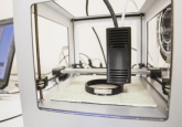
The goal of this editorial is to provide a brief overview of the process of 3D printing as well as highlighting the utilization of 3D models of congenital heart disease (CHD) when explaining diagnoses to patients and their families, teaching trainees about CHD and in pre-procedural planning for both interventional cardiologists as well as cardiac surgeons. Three-dimensional (3D) printing was initially developed in the 1980s with subsequent gains in the field of medicine as well as improvements in consumer use. The process for printing a 3D model typically involves an image dataset, in medicine from computed tomography (CT) or magnetic resonance (MR) data. The areas to be printed are selected and highlighted, undergoing a process known as segmentation [1]. Although there...