Bioprinting: the future of medicine
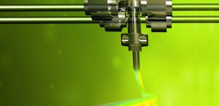
This editorial discusses the history, promise and challenges associated with bioprinting, an enabling biofabrication technology that is anticipated to transform the future of medicine.
Yu Shrike Zhang1
1Division of Engineering in Medicine, Department of Medicine, Brigham and Women’s Hospital, Harvard Medical School, Cambridge, MA 02139, USA; [email protected]
While 3D printing has long been adapted for use in many areas such as home, art and education since its invention 30 years ago [1], its combination with biological materials, or bioprinting, has been present for merely a decade or so [2,3]. Although relatively new, this capable technology is already starting to revolutionize the field of medicine by providing unprecedented flexibility and fidelity in creating volumetric biological structures that mimic those of their native counterparts in the human body [4].
Compared with conventional biofabrication technologies, the bioprinting process is highly reproducible due to its programmed robotic controls, and, for the same reason, large-scale automation is also made possible. As such, 3D bioprinting has already found extensive applications in both engineering functional tissue constructs for regeneration of injuries in vivo [5] and generating biomimetic organoids for predictive screening of biomolecules in vitro [6].
Nevertheless, several challenges have so far limited effective translation of bioprinting, most pressing ones are the need for optimizations of the biological materials, including both the bioinks and the cells, and the multimaterial capacity to simultaneously integrate several types of biological materials.
The cells
The cells are a critical component in achieving successful bioprinting of functional tissue constructs. Conventionally, somatic cells that are terminally differentiated have been the main source of cells. With the development of the stem cell technologies, it has become increasingly popular to use these cells, such as mesenchymal stem cells, amniotic fluid-derived stem cells and induced pluripotent stem cells, or their derivative cell types, as the cells for bioprinting [7].
Besides the almost unlimited sources, these cells may be further personalized in a patient-specific manner. While multi- or pluripotent, this unique property of the stem cells simultaneously hinders their applications, mostly due to concerns associated with the undifferentiated population of the cells that are prone to cause unwanted side effects. Therefore, further optimization of methods (e.g. CRISPR [8,9]) that can tightly control the stem cell fate is anticipated to significantly improve the use of these cells for translational bioprinting.
The bioink
The bioink refers to the materials, usually in the hydrogel form, either used alone or for carrying the embedded cells during the bioprinting process to serve as the supporting matrix [10-12]. In a sense, the bioinks function as the substitutes for the extracellular matrix to maximally ensure proper microenvironments where cells can reside and respond in a similar way they behave in their native niches. The design of the bioinks is thus critical, especially when cells are encapsulated within the bioinks.
First, the biomaterials should be cytocompatible not exerting any toxicity to the cells, either embedded or post-seeded, beyond a level suitable for desired applications. Second, the bioprinting conditions ought to be benign to avoid potential damage (e.g. overly high shear stresses in the case of extrusion bioprinting [13]) to the encapsulated cells during the bioprinting processes. Third, the crosslinking parameters need to not cause cell malfunctions, such as the possible adoption of visible light photoinitiators [14] when photocrosslinkable bioinks are involved in order to avoid exposures to ultraviolet light.
Multimaterial capacity
Bioprinting of tissue constructs at clinically relevant scales has been restricted by the scalability most bioprinting systems lack. A microfluidic tree-structured printhead design possessing parallel nozzles [15] may greatly enhance the throughput of bioprinting with optimized parameters. Alternatively, aggregates of cells may be used as the bioink, providing larger tissue building units to increase the bioprinting efficiency [16].
Another concern is the complexity bioprinting systems can reach in generating tissue constructs. In the nature, biological tissues are highly complex containing hierarchically assembled structures assembled across multiple length scales, which cannot be realized using single material bioprinters. To this end, several extrusion bioprinter designs that allow for sequential or simultaneous deposition of multiple bioinks from several nozzles that are mechanically co-registered during a bioprinting session have been developed [17,18].
More recently, a single nozzle, multichannel printhead design has also been reported to enable digitally tunable multibioink deposition, leading to a >10-time improvement in bioprinting speed compared with multinozzle systems [19]. Similarly, multimaterial stereolithographic bioprinters have demonstrated capability to construct hierarchical tissues as well [20]. While each platform has its own pros and cons, the integration of complementary multimaterial and high-throughput capacities may make more powerful bioprinting systems.
Conclusions
While advances have been made in attempt to meet the pressing needs in 3D bioprinting including those related with cells, bioinks and multimaterial capacity, careful optimizations of various parameters are application-specific. With further development of strategies overcoming the challenges associated with bioprinting, it is anticipated that this enabling biofabrication technology will eventually achieve bioprinting of clinically relevant human tissues and transform the future of medicine.
Acknowledgements
- The author acknowledges supports from the National Cancer Institute of the National Institutes of Health Pathway to Independence Award (K99CA201603) and the LUSH Prize.
Financial & competing interests disclosure
- The author has no relevant affiliations or financial involvement with any organization or entity with a financial interest in or financial conflict with the subject matter or materials discussed in the manuscript. This includes employment, consultancies, honoraria, stock ownership or options, expert testimony, grants or patents received or pending, or royalties. No writing assistance was utilized in the production of this manuscript.
References
- Hull CW. Apparatus for production of three-dimensional objects by stereolithography. (Ed.^(Eds) (Google Patents, 1986)
- Boland T, Mironov V, Gutowska A, Roth E, Markwald RR. Cell and organ printing 2: Fusion of cell aggregates in threedimensional gels. The Anatomical Record Part A: discoveries in molecular, cellular, and evolutionary biology, 272(2), 497-502 (2003).
- Wilson WC, Boland T. Cell and organ printing 1: protein and cell printers. The Anatomical Record, 272(2), 491-496 (2003).
- Zhang YS, Yue K, Aleman J et al. 3D Bioprinting for Tissue and Organ Fabrication. Ann. Biomed. Eng., 45(1), 148-163 (2017).
- Murphy SV, Atala A. 3D bioprinting of tissues and organs. Nat. Biotechnol., 32(8), 773-785 (2014).
- Peng W, Unutmaz D, Ozbolat IT. Bioprinting towards physiologically relevant tissue models for pharmaceutics. Trends Biotechnol., 34(9), 722-732 (2016).
- Tasoglu S, Demirci U. Bioprinting for stem cell research. Trends Biotechnol., 31(1), 10-19 (2013).
- Makarova KS, Zhang F, Koonin EV. SnapShot: Class 2 CRISPR-Cas Systems. Cell, 168(1—2), 328-328.e321 (2017).
- Makarova KS, Zhang F, Koonin EV. SnapShot: Class 1 CRISPR-Cas Systems. Cell, 168(5), 946-946.e941 (2017).
- Jungst T, Smolan W, Schacht K, Scheibel T, Groll Jr. Strategies and Molecular Design Criteria for 3D Printable Hydrogels. Chem. Rev.,(2015).
- Chimene D, Lennox KK, Kaunas RR, Gaharwar AK. Advanced Bioinks for 3D Printing: A Materials Science Perspective. Ann. Biomed. Eng., 1-13 (2016).
- Hospodiuk M, Dey M, Sosnoski D, Ozbolat IT. The bioink: A comprehensive review on bioprintable materials. Biotechnol. Adv.,(2017).
- Blaeser A, Duarte Campos DF, Puster U, Richtering W, Stevens MM, Fischer H. Controlling Shear Stress in 3D Bioprinting is a Key Factor to Balance Printing Resolution and Stem Cell Integrity. Adv. Healthcare Mater., 5(3), 326-333 (2016).
- Xiao P, Zhang J, Dumur F et al. Visible light sensitive photoinitiating systems: Recent progress in cationic and radical photopolymerization reactions under soft conditions. Prog. Polym. Sci., 41, 32-66 (2015).
- Hansen CJ, Saksena R, Kolesky DB et al. HighThroughput Printing via Microvascular Multinozzle Arrays. Adv. Mater., 25(1), 96-102 (2013).
- Mironov V, Visconti RP, Kasyanov V, Forgacs G, Drake CJ, Markwald RR. Organ printing: tissue spheroids as building blocks. Biomaterials, 30(12), 2164-2174 (2009).
- Kolesky DB, Truby RL, Gladman AS, Busbee TA, Homan KA, Lewis JA. 3D Bioprinting of Vascularized, Heterogeneous Cell-Laden Tissue Constructs. Adv. Mater., 26(19), 3124-3130 (2014).
- Kang H-W, Lee SJ, Ko IK, Kengla C, Yoo JJ, Atala A. A 3D bioprinting system to produce human-scale tissue constructs with structural integrity. Nat. Biotechnol., 34(3), 312-319 (2016).
- Liu W, Zhang YS, Heinrich MA et al. Rapid Continuous Multimaterial Extrusion Bioprinting. Adv. Mater., 29, 1604630 (2017).
- Ma X, Qu X, Zhu W et al. Deterministically patterned biomimetic human iPSC-derived hepatic model via rapid 3D bioprinting. Proct. Natl. Acad. Sci. U.S.A., 113(8), 2206-2211 (2016).
