Editorial: biomaterial choice is crucial to effective iPSC laser bioprinting
In this article, Geoffrey Potjewyd (University of Manchester, UK) reviews a recent report from Biofabrication.
Geoffrey Potjewyd1
1Regenerative Medicine & Neuroscience PhD student, University of Manchester (UK)
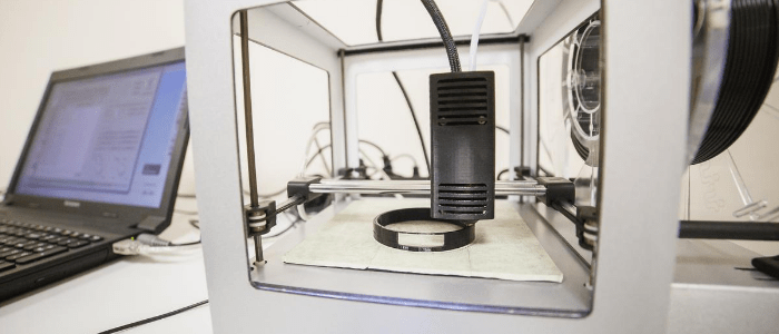
3D printing is increasingly being utilized in medical communities, with the rapid and consistent printing being used to develop prosthetics, medical devices, and even mock surgical tissues. The application of 3D printing to ’bioprint‘ biological material is one which is being utilized more and more in recent years, with the promise of creating better in vitro models for research as well as creating tissues and organs for transplantation.
This has been aided in part by the development of stem cell research, and in particular induced pluripotent stem cells (iPSCs). These cells can be reprogrammed from human adult stem cells which are extracted in a simple skin biopsy. From there, cells can be differentiated into the different cell types of a tissue and combined to create a multi-cellular tissue.
The combination of 3D bioprinting and iPSCs allows for cells to be printed into precise spatiotemporal locations within a tissue construct to build human 3D tissues and organs in the lab as well as printed patterns for high throughput screening or network development. The use of patient stem cells enables a stratified, patient specific tissue model for research, or development of transplantable tissues and organs.
The combination of bioprinting and stem cells is not simple, however, with many different obstacles to be considered before successful tissue engineering can occur. In order to maintain a 3D structure for cells, they must be encapsulated within a biomaterial which provides the 3D extracellular environment native to the tissue type. This requires polymer chemistry to create a biomaterial which is printable (a bioink), is able to house the cells for 3D cell culture after printing (biocompatible), and also provides the necessary biophysical and biochemical environment for cells to elicit their native responses. In addition to this, iPSCs are notoriously sensitive to different conditions and the stresses imposed by the handling of cells make bioprinting potentially demanding.
A research group from Hannover (Germany) has investigated the effect of using different biomaterials and the physical process that printing has upon iPSCs, with specific insight into how this affects the iPSC survival, pluripotency and ability to differentiate.
In particular the group has focused on the effect of laser bioprinting on cells. This is a bioprinting technique that does not require the extrusion of cells through a nozzle or needle, so does not impose the physical shear stress that is commonplace with extrusion based bioprinting techniques.
The researchers found that cell viability was decreased slightly following printing, but cell membrane integrity and metabolic activity of printed cells was not affected. The decrease in cell viability was isolated to shortly after printing, with cell death being the same in all the latter time points.
The surface which cells were printed on was also investigated, with different surface coatings altering cell behavior. From this they found that Matrigel was optimal to avoid cell aggregation as the other gel coatings promoted aggregation and clumping of cells. In addition to this, the Matrigel coating also supported the patterned deposition from the laser printing.
The group established that using a hyaluronic acid bioink aided maintenance of pluripotency post-printing, with iPSCs being differentiated into all three germ layers. Importantly this was also independent of printing, meaning the printing procedure did not affect the differentiation capability.
Overall the group has found that printing human iPSCs does not affect the cells negatively, although the bioinks used can have a significant effect on cells after printing. In particular the researchers found that laser printing using a hyaluronic acid bioink mixed with iPSC media onto Matrigel had the best results, with defined printing as well as maintaining viability, pluripotency and differentiation potential.
This research builds on previous laser bioprinting work that has shown that immortalized, primary and stem cell lines can be printed without adversely affecting cell survival or behavior. Authors state that to their knowledge this is the first example of laser bioprinting of iPSCs, and could lead to applications of the technique in disease modeling and innovative in vitro assays.
Source: Koch L, Deiwick A, Franke A, Schwanke A, Haverich A, Zweigerdt R and Chichkov BN.Laser bioprinting of human induced pluripotent stem cells — the effect of printing and biomaterials on cell survival, pluripotency, and differentiation. Biofabrication. https://doi.org/10.1088/1758-5090/aab981. (2018) (Epub ahead of print)