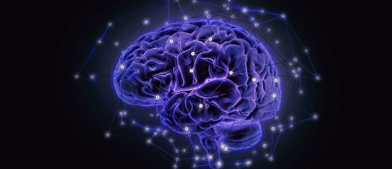
Mind mapping: modern research method allows for the study of brain processes associated with disease
Researchers have designed a study approach that enables a substantially more detailed analysis of the brain processes at work in certain neurological illnesses. This is accomplished through developing human cortical organoids and implanting them into developing mouse brains to observe how they integrate and operate over time. The study was funded and supported by the National Institute of Mental Health (MD, USA), a division of the National Institutes of Health (MD, USA), and appears in Nature.
“This work provides a significant advance in the ability of scientists to study the cellular and circuit underpinnings of complex human brain disorders. It allows organoids to get ‘wired’ in a more biologically relevant context and function in ways they can’t do in a petri dish,” said David Panchision, chief of the Developmental and Genomic Neuroscience Research Branch in the Division of Neuroscience and Basic Behavioral Science at the National Institute of Mental Health.
Sergiu Pasca and colleagues at Stanford University (CA, USA) have shown that a cortical organoid produced from human stem cells can be grafted onto and integrated into a developing rat brain to better understand specific developmental and physiological processes. The results indicate that transplanted organoids could be an effective model for examining disease development pathways.
Cortical organoids are three-dimensional cultures of human stem cells that can mimic several of the developmental processes observed in ordinary brains. They are commonly used as a model by researchers to investigate how certain components of the human brain grow and function. However, cortical organoids, lack intrinsic connection inherent in average healthy human brains, which restricts their utility in understanding complex brain processes. Scientists have attempted to circumvent many of these restrictions by implanting individual human neurons into the brains of adult rodents. The transplanted neurons connect to the rodent brain cells, but because of the developmental limitations of the adult rat brain, they do not fully integrate with rodent brain cells.
In the present study, the researchers pioneered the use of brain organoids for research by transplanting a fully formed human cortical organoid into a maturing rat brain. This technique generates a unit of human tissue that can be easily manipulated and analyzed. Human-induced pluripotent stem cells were used to produce the cortical organoids, which were previously pioneered in Sergiu Pasca’s lab. The organoids were subsequently transplanted into the rat primary somatosensory cortex, the region of the brain responsible for the processing of sensation.
The researchers found no impairments in motor or cognitive function or brain activity in the rats that received the transplanted organoid. The transplanted tissue was nourished via blood arteries from the rat brain, which had developed over time.
The scientists infected a cortical organoid with a viral tracer which travels across brain cells as a marker of functional connections. This allows the determination of the extent to which the organoids may integrate into the rat somatosensory cortex. The viral tracer was detected in several areas of the brain including the ventrobasal nucleus and the somatosensory cortex, after transplanting the labeled organoid onto the rat’s main somatosensory cortex.
Furthermore, the researchers discovered new links between the thalamus and the transplanted region. Electrical stimulation and stimulation of the rat’s whiskers were used to activate these connections, demonstrating that they were experiencing the relevant sensory input. Furthermore, the researchers were able to achieve regulation of the rat’s reward-seeking behaviors by activating human neurons in the transplanted organoid. The evidence points to the transplanted organoid’s functional integration into certain brain networks.
Following seven to eight months of development, the transplanted brain organoid mirrored neurons from human brain tissue more so than human organoids grown in cell culture. The researchers questioned if they could employ transplanted organoids to study parts of human disease processes since transplanted organoids resembled the anatomical and functional characteristics of human cortical neurons so distinctly.
“The promise of this platform is not only in identifying what molecular processes underlie the advanced maturation of human neurons in living circuits and leveraging it to improve conventional in vitro models, but also in providing behavioral readouts for human neurons,” said Dr. Pasca.
In order to investigate this, cortical organoids were developed from three persons with Timothy syndrome, a rare genetic illness associated with autism and epilepsy, and three other participants without any known disorders, and transplanted them into the rat brain. Both types of organoids integrated into the rat somatosensory cortex, but Timothy Syndrome organoids displayed structural abnormalities. These same structural abnormalities were not present in organoids made from Timothy Syndrome patients’ cells and grown in cell culture.
“These experiments suggest that this novel approach can capture processes that go beyond what we can detect with current in vitro models,” said Dr. Pasca. “This is important because many of the changes that cause psychiatric disease are likely subtle differences at the circuit level.
Source: https://www.sciencedaily.com/releases/2022/10/221012132345.htm