Brain organoids reveal new treatment strategy for herpes simplex encephalitis
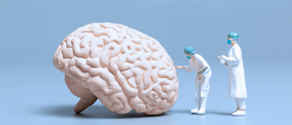
A critical pathway in the pathogenesis of herpes simplex encephalitis has been revealed using brain organoids, enabling the identification of improved treatment paradigms.
A recent study, emanating from the Max Delbrück Center’s Organoid Platform (Berlin, Germany) and led by senior authors Nikolaus Rajewsky and Markus Landthaler, has utilized brain organoids to identify a treatment regimen for herpes simplex encephalitis that could improve patient outcomes. This development could have a dramatic impact on outcomes for viral encephalitis patients, reducing the number and severity of their neurological sequela.
Two thirds of humans are infected with the virus herpes simplex virus-1 (HSV-1), initially replicating in epithelial cells before integrating into trigeminal ganglia cells, where it resides in the neurons, inactive and benign until a stressor activates it, typically leading to a cold sore. In 5-15% of infectious encephalitis cases, the infection spreads up the nerves to the brain, leading to a life-threatening condition. The current standard of care involves the use of the antiviral acyclovir. While this treatment dramatically reduces mortality, patients are often left with neurological sequelae, such as memory loss and seizures.
The study of encephalitis caused by HSV-1 can be challenging; the virus only infects humans, so animal models to study the condition have serious limitations, and obtaining human neural tissue is marred with ethical and practical difficulty.
So, how do you study a condition that is specific to human tissue and identify a therapeutic solution that improves the quality of life post recovery for patients? Agnieszka Rybak-Wolf, first author and Head of the Organoid Technology Platform at the Max Delbrück Center, set to work to answer this question and generated 0.5 cm wide brain organoids from induced pluripotent stem cells that “look a bit like small clouds of tissue.”
The team then infected these organoids with a green fluorescent protein (GFP) expressing the HSV-1 strain. Immunofluorescence imaging techniques and spatial transcriptomic profiling enabled the team to detect the GFP and mRNA respectively and track the spread of the virus through the organoids. After infection, single-cell RNA sequencing was used to investigate transcriptional changes and the molecular pathways active in the organoids, while calcium imaging provided information on neuronal activity.
This approach allowed them to identify that the TNF-α signaling pathway was significantly upregulated during infection. Furthermore, when acyclovir was administered, HSV-1 stopped reproducing, but tissue damage continued, with subsequent investigations revealing that the TNF-α pathway was still active during this period.
Commenting on the finding, co-first author Tancredi Pentimalli explained that “the inflammation pathway is a key natural defense to the virus but when we block viral replication with anti-viral drugs, the overzealous inflammatory response could instead become damaging.”
Following up on this finding, the team tested the infected organoid’s response to both antivirals and anti-inflammatories, necrostatin-1 or bardoxolone methyl, that antagonize the TNF-α pathway. With this combination, the team observed significant protection of the brain organoids from the typical injury caused by infection.
Pentimalli concluded, “I hope that clinical investigators will set up clinical trials evaluating the efficacy of new anti-viral and anti-inflammatory combination therapies in herpes encephalitis patients, ultimately translating our findings from the bench to the bed side.”