Innovative aMace technology for complex pelvic tumor resection and reconstruction

New, 3D-printed hip implants have made five bone cancer patients, two clinical teams and Materialise, very happy. Together with the orthopaedic departments of the University Children’s Hospital Basel (UKBB), Switzerland and the Righospitalet in Copenhagen, Denmark, Materialise participated in a study to reconstruct periacetabular defects caused by tumors. Dr. Krieg and his colleagues shared their findings in the medical magazine, Leading Opinions.
New, 3D-printed hip implants have made five bone cancer patients, two clinical teams and Materialise, very happy. Together with the orthopaedic departments of the University Children’s Hospital Basel (UKBB), Switzerland and the Righospitalet in Copenhagen, Denmark, Materialise participated in a study to reconstruct periacetabular defects caused by tumors. Dr. Krieg and his colleagues shared their findings in the medical magazine, Leading Opinions.
Periacetabular reconstruction after tumor resection
Approximately 8-20% of all bone tumors occur in the pelvis, one third of which are malignant. For these challenging bone defects particularly, a patient-specific solution is necessary. After three years of anatomical examinations, biomechanical experiments, further analysis of modularity and one-time surgeries, innovation prevailed.
Thanks to Materialise aMace technology, the orthopaedic teams turned what were very challenging bone defects into successful periacetabular reconstructions. With the support of biomedical engineers and both clinical groups from Copenhagen, Denmark and Basel Switzerland, the new aMace Tumor Reconstruction System was tested in five patients with periacetabular defects.
Five successful surgeries
For two of the patients, a two-step procedure of the aMace implant initially was performed. In association with Materialise, the medical teams planned the surgery with 3D virtual imaging, starting with MRI and CT scans of the tumor. Then, the tumor was removed using patient-specific resection guides. Ten days later, based on a CT scan of the resected bone, the final aMace and custom plates were designed and 3D printed, and the surgical team performed the final surgery. In the three other cases, a one-time procedure with direct aMace implantation was performed thanks to patient specific cutting guides.
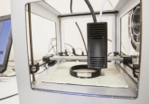
Left to right: Scaffold with modular plates and screws, patient-specific resection guides, test models, patient-specific drilling guides, bone model
Read more: http://www.materialise.com/en/blog/pelvic-reconstruction-3D