Lighting the way: how PISA is revolutionizing 3D-printed tissues

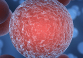
A new light-based 3D-printing method has been developed, providing a more efficient process for creating organs-on-a-chip.
Researchers from the Missouri University of Science and Technology (Missouri S&T; MO, USA) have reimagined the traditional method for creating 3D printing tissues for organs-on-a-chip applications to address a major challenge in tissue engineering.
Organs-on-a-chip are small, tissue-like devices designed to study how human tissues respond to new medicines or treatments without the need for animal or human testing. Traditionally, these devices are fabricated point by point technique, which involves dispensing individual droplets of bioink from a print head onto a surface to construct structures layer by layer. While this method allows for precise placement of materials, it is both time-consuming and expensive, particularly when creating the intricate capillary networks that tissues rely on for survival.
“Re-creating those dense microcapillary networks is a major engineering challenge for tissue engineering,” expressed Anthony Convertine, an associate professor of materials science and engineering.
 Bridging the gap: 3D-printed scaffolds create new path for spinal cord repair
A novel technique uses microscopic printed channels to guide stem cells across spinal cord injuries, restoring connections once thought permanently lost.
To overcome this challenge and improve the efficiency of 3D printing tissues, Convertine and his team developed a novel method called polymerization-induced self-assembly (PISA) printing. This approach uses a light-curable, self-assembling resin to form sacrificial structures. PISA printing combines two key technologies: reversible addition-fragmentation chain transfer (RAFT) polymerization, which controls the building of polymer chains, and digital light projection (DLP) photolithography, a light-based 3D printing technique that cures materials layer by layer using projected patterns. Unlike traditional 3D printing, which relies on permanent covalent crosslinks, PISA printing creates structures held together by physical networks – temporary sacrificial structures that can be dissolved after printing.
“After printing, we dissolve those [sacrificial] structures to leave clean, precise microchannels. It is faster, simpler and easier to scale,” explained Convertine.
The team also developed a simplified “one-pot” method to create multi-chain transfer agent scaffolds, which act as molecular frameworks to guide polymer organization. These scaffolds form strong structures through “interparticle bridges and knots,” allowing polymer chains to wrap around and through each other.
The researchers demonstrated the success of their technique by creating tiny, blood vessel-like channels that fluids can flow through, effectively mimicking the structure and function of capillaries – an essential element for tissue survival.
This method integrates the chip’s entire microchannel system into a single mixture, significantly reducing processing steps and offering a faster, more efficient, and adaptable platform for microfabrication, rapid prototyping, and advanced tissue engineering applications, including the streamlined prototyping and testing of designs.