Top tips for incorporating organoids into your research: an interview with Elfie Rödel
Dr. Elfie Rödel is a Project Manager in the Development Department of PromoCell (Heidelberg, Germany) where she has worked for almost 10 years, exploring the development and optimization of cell culture products and special product applications, such as 3D culturing. With a primary focus on epithelial cells Elfie is working to reduce the use of animal-derived components or serum factors in culture medium and improving the utility and practicality of implementing organoids and complex cell models in research pipelines.
Here, Elfie discusses selecting appropriate 3D cultures for different applications, the challenges of establishing and using these models and shares her best practice tips for working with these exciting new cultures.
This interview is part of the RegMedNet In Focus on growing organoids. Discover expert opinions on this topic by visiting our feature homepage.
What factors require consideration when selecting an appropriate 3D culture system for specific applications?
For many decades, the cultivation of isolated primary cells from human tissue as a so-called “submerged culture” was the gold standard in classical cell culture. However, cell cultivation in 2D carries the risk that the functionality of the cells, which is significantly influenced by the tissue structures occurring in vivo, is not fully developed and artifacts can occur in the experiment. Researchers then developed systems for the 3D culture of cells, generating cultures that better reflect the complexity of tissues or organs.
There are 3D culture systems of various epithelial tissues, such as the liver, the colon or the airways. Before selecting the 3D culture system, it must be theoretically questioned and tested in the design of the experiment: Is the 3D culture system suitable for answering my specific question? In addition, the 3D culture system must be scientifically recognized and accepted.
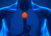
Airway epithelial cells are an excellent example of these cultures and their use. Even before the COVID-19 pandemic, 3D cultivation of airway epithelial cells was a popular method. It is used for research on the basic mechanisms and possible therapeutic approaches for respiratory diseases that are increasing worldwide, such as asthma or COPD. Airway epithelial cells form a barrier function between the tissue of the respiratory tract and the inflowing ambient air through the mouth and nose area. To mimic this situation in vitro, epithelial cells were cultured on a porous membrane that allowed access to the culture medium on the basal side and access to ambient air on the apical side of the cell. This is often referred to as an air-liquid interface (ALI) culture. Under these culture conditions, the cells form a measurable epithelial barrier function between the two compartments. This functionality can be demonstrated with a voltohmmeter, for example, and can be used for various approaches such as cell-virus interactions, cytotoxicity tests or drug screening.
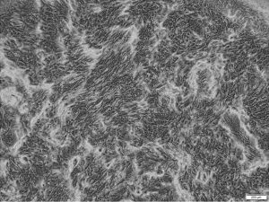
After 4 weeks of airlift culture HBEpC show typical morphology and high viability.
However, the ALI culture has the disadvantage that it is not suitable for high-throughput tests due to the dependence on multiwell inserts with porous membranes. For this purpose, the use of airway organoids would be recommended, as these can be cultivated in 96-well plates in an extracellular matrix (ECM) without any problems. However, the production of airway organoids requires a little more finesse compared to the ALI culture, although the cell starting material is the same. When establishing an organoid protocol in your own laboratory, it is therefore a great time saver to use prescreened products.
How can researchers select the most suitable cell culture material for producing their own organoids?
The procurement of cell culture materials is not always easy, especially in times of global material shortages. The important materials in 3D culture include the following products: cells, cell-culture medium, cell-culture plastic and an ECM. All materials should be as robust, deliverable, easy to use and QC-qualified as possible.
Regarding the cell culture medium, a distinction is made between an expansion medium, phase I, and a differentiation medium, phase II. There are numerous peer-reviewed publications on the production of 3D cultures that contain detailed step-by-step instructions. This includes manufacturing protocols for expansion and differentiation media. It should be noted that a complete cell-culture medium – consisting of amino acids, salts, trace elements, lipids, growth factors and cytokines, among other things – contains more than 50 components and is therefore prone to error in production. In order to eliminate as many sources of error as possible from the already variable cell culture model, it is advisable to use commercial products.
Also, many laboratories do not have access to tissue material to isolate primary cells. Using the scientific literature can help to identify commercial products that have already been used successfully.
Do you have any best practices to share for moving to and working with 3D cell cultures, e.g. airway organoids?
If you already have experience in expanding primary cells, the step to 3D cell culture is not a big one. Every 3D culture begins with an expansion of the cells in 2D. For airway organoids, primary human bronchial epithelial cells can be used for this purpose. It should be noted that primary cells have a finite lifespan. With increasing population doubling, the ability of cells to differentiate can decrease. This must be considered if you want to differentiate the cells later. For the 3D cultivation of bronchial epithelial cells, we have developed a robust air-liquid interface medium that is qualified for a long-lasting, measurable barrier function over a period of at least 4 weeks. We found that this medium was not only suitable for a functional culture on porous membranes, but also for a functional 3D culture in an ECM.
Both applications were developed according to best practice guidelines. We were able to gain a lot of experience during the establishment of both protocols. When working with biological systems, the susceptibility to failure is very high. It is therefore important to use as many robust constants as possible. Not every tissue donation is suitable for generating bronchial epithelial cells capable of differentiation. In order to qualify cell batches for the 3D cell culture, cell batches need to be screened in a time-consuming manner. Other possible confounding factors include cell culture plastic and ECM.
In the case of cell culture plastic, it must be said that with organoid technology it is essential that the plastic has non-adhesive properties, otherwise the cells may adhere to the plastic surface and not remain in suspension. If you want to perform high-throughput experiments on your 3D organoid cultures, 96-well plates with U-bottom are recommended. Even with small wells, the cells can be well distributed in the ECM and are easy to examine under the microscope.
Various ECM variants for organoids can be found in the literature, with commercial basement membrane matrix occurring frequently. With ECM we have had good experiences using commercial basal membrane extract obtained from Engelbreth-Holm-Swarm (EHS) mouse sarcoma cells. In addition to the numerous adhesive proteins, this matrix also contains growth factors such as TGF-ß or EGF. These growth factors are known regulators of proliferation and differentiation. A growth-factor-reduced basal membrane extract should therefore be used to prevent possible suppression of differentiation.
It should also be noted that there are different orientations for the airway organoids. With airway organoids, a distinction is made between apical-in and apical-out models. This describes the orientation of the polarized epithelial cell layer, which can be very well distinguished from one another microscopically. The ciliated cells point outwards in the apical-out model. This has the advantage that these cells are freely accessible for experiments. For example, in order to stimulate the polarized epithelial cells, one does not need to microinject for example, virus material or mechanical fragmentation of the apical-in models. It is already known that the orientation of cell polarity can be influenced by the ECM.
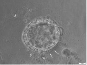
Differentiation of HBEpC after 16 days of 3D culture in ALI-Airway. Bright-field microscopy shows the polarized epithelial cell lining of the organoids and the inner central lumen. Arrows indicate differentiated, outside oriented ciliated cells. Scale bar = 20 µm.
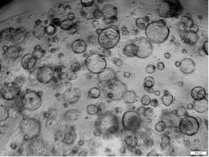
Airway organoids with self-renewal potential can be used for longterm culture.
From a technical point of view apical-in airway organoids can be detached from the ECM and then triggered to apical-out in an ECM-free suspension culture. However, a disadvantage of this structural organoid switch is that many organoids lose their integrity and, for example, fuse with other organoids. With our Application Note, we have succeeded in creating a simple protocol for the 3D cultivation of bronchial epithelial cells that favors the generation of apical-out models.
What are the benefits of working with organoids developed from healthy human primary cells versus cell lines?
An organoid requires cells that are capable of differentiation and have the potential to self-assemble into a specific structure. Cell lines are usually isolated cells from tumors or metastatic tissue of patients. Such samples are collected for biobanking and can be used for applications such as cancer research.
An interesting aspect here is the possibility of drug screening for personalized medicine. However, only a few patient-derived cells can be used spontaneously for 3D culture as so-called tumorspheres. Most cell preparations require a 3D culture medium with a specific cocktail to activate or inhibit signaling pathways to form a functional 3D structure.
A disadvantage compared to healthy cells is that there are major differences between the growth potential and growth rate of tumor cells. The genetic instability of tumor cells results in large fluctuations, which can lead to a reduction in robust data sets. Another point is that during the preparation of tumor cells, cells from the surrounding healthy tissue can also be isolated and afterwards contaminate the pure tumor cell population. There is a risk here that these contaminating cells can overgrow the tumor culture. The organoid media were originally developed for epithelial cells and could therefore favor the overgrowth with healthy epithelial cells.
The primary cells from healthy patient material have the advantage that they are usually suitable for 3D culture over a longer period of time. So-called prescreened cells also offer the advantage that they have been tested regarding their cell identity, HLA-type and growth characteristics and have been subjected to a functional test regarding the barrier function. This is the basis for generating robust assays.
What are the greatest challenges faced in the production of respiratory organoids and how can these be overcome?
The biggest challenge with organoids is that this model lacks the influence of an intact tissue structure. A complex tissue supports structure and an interacting environment is essential for building fully developed functionality with tissue retention.
In addition to the epithelium, natural respiratory tissue also contains smooth muscle cells, fibroblasts, and an ECM-rich stroma. It is known that numerous respiratory diseases can lead to long-lasting inflammatory reactions in the tissue and thus permanent tissue remodeling. These structural changes in the layer of smooth muscle cells can lead to a fibrotic change and thus also to a reduction in the lumen and the associated airflow. These structural cells do not occur in the airway organoid culture. Researchers using ALI cultures have so far succeeded in co-cultivating airway epithelial cells with fibroblasts and endothelial cells.
Another point is the lack of immune cells. The lining epithelial cell layer is part of the immune system and is in contact with the immune cells. The immune cells are another protective shield of the epithelial barrier. For example, macrophages, eosinophils, mast cells or neutrophils are responsible for the recognition and elimination of pathogens. It has been observed not only with pathogens, but also with structural changes that immune cells migrate into the diseased tissue. In COPD patients, for example, there are increased amounts of T-lymphocytes in the tissue. These important cell-cell interactions are missing in the current airway organoid model.
However, there are some efforts to co-culture airway organoids with macrophages. First results are promising. The natural “macrophage clearance” could be simulated by the co-culture. The inclusion of these immunological aspects can also play an important role in the development of new drugs.
Disclaimer
The opinions expressed in this interview are those of the interviewee and do not necessarily reflect the views of RegMedNet or Future Science Group.
In association with
![]()