Q&A highlights: 3D cell culture: embracing the 3Rs for clinically-relevant results
The increasing use of physiologically-relevant 3D cell cultures such as PeptiGel® is rapidly advancing pre-clinical studies, due to their ability to mimic the in vivo environment and produce clinically relevant data. Robust use of 3D cultures makes them compliant with the 3Rs: “replacement, reduction and refinement” minimizing animal use in research.
In a recent webinar, Aline Miller, CEO and Professor, Manchester BIOGEL and The University of Manchester (both UK), discussed recent developments in 3D cell cultures and their application in regenerative medicine and drug discovery.
Watch the webinar on-demand now or read on for highlights of the Q&A session following Aline’s presentation.
Jump to:
About PeptiGels®
Is the hydrogel morphology similar to nanofibers or any nanostructures?
Yes, our peptide hydrogels are composed of a beta-sheet rich fibrillar structure, reminiscent of natural extracellular matrix.
Are your gels physically or chemically crosslinked? If physical, do they wash away over time? If chemical, how do/would they degrade in vivo?
Our hydrogel products are physically crosslinked and do degrade slowly over time, the kinetics of which depends on their surrounding environment and how careful users are when changing media. We regularly undertake culture experiments for 4-6 weeks and have evidence of PeptiGels® surviving at injection sites in vivo for at least up to 2 months.
Can you comment on the dependence of the rheology/injectability of the gel(s) with temperature?
PeptiGels® are shear-thinning and injectable. PeptiGels® are not dependent on temperature within the range of 4-50oC and are ready to use at room temperature.
How stable are the peptide hydrogels?
On average, most of our hydrogels have been used for in vitro cultures lasting 4–6 weeks. They are also suitable for longer cultures. In vivo studies undertaken to date show that the peptide hydrogels are stable for 2-6 weeks, depending on the environment of the injection site. Our technical team can advise you if you are planning longer experiments.
Is there any information on the thickness of the RGD and GFOGER gels?
We have a catalogue of different functionalized gels, including RGD and GFOGER functionalized PeptiGels®, and we provide all the necessary technical information and details on how to set up your experiments for each product.
Please visit our site www.manchesterbiogel.com for more information.
How does charge affect the cell morphology and cell communication?
Our PeptiGels® have neutral or positive charge in culture as all cells like a different environment. It is well known that a positive charge typically encourages cell adhesion. However, we can supply gels at certain charges based on customer request.
What are the advantages of scaffold-based 3D culture over other 3D culture methods?
There are several advantages of using scaffold-based 3D culture such as PeptiGels® over other 3D culture methods. These include giving a more representative in vivo environment to allow the generation of more physiologically and clinically relevant data. Synthetic scaffolds, such as our PeptiGels®, can be fine-tuned mechanically and functionally allowing the creation of an in vitro environment that mimics the in vivo counterpart, and essentially any human tissue. These benefits, alongside the potential scalability and tunability open the door to new and exciting applications.
To learn more, visit our website www.manchesterbiogel.com or [email protected].
Working with PeptiGel®
How do you ensure the materials you use give you the same results every time?
PeptiGels® are fully synthetic and produced from fully defined formulations, hence there is no batch-to-batch variability ensuring results are reproducible and reliable every time.
Have you found a limit to the thickness of your 3D cell culture? Can you vascularize the gels?
The thickness we recommend is 1.05mm with a well’s surface area of 1.91mm2 on our previous experience. There will be a limit on the thickness of PeptiGels® due to diffusion of nutrients and cell waste products, but this is not something we have explored as yet. Yes, we can promote vascularization by incorporating appropriate cell recognition sequences.
How do you incorporate functionality within the peptide hydrogels? And how can you control their concentrations?
Functionality is incorporated into our peptide hydrogels with the addition of protein mimetics tagged to the end of our self-assembling peptides. We control their concentration at the production stage and can vary these based on customer’s requests if desired. Functionality can also be done manually by customers through the addition of protein factors into the culture medium before adding to the hydrogels.
Can you make scaffolds that are multifunctional?
Yes. Any combination desired is possible and can be varied systematically.
How do you know which hydrogel to use for each cell type?
We have a reference guide to recommend which hydrogel is suitable for each cell type. Our experienced technical team can also guide you in the selection of your hydrogel. Please get in touch at [email protected]
How do you extract cells from the hydrogels to do things like qPCR?
We have detailed protocols to guide you in doing certain experiments such as qPCR. Please check our protocols on our website www.manchesterbiogel.com or get in touch at [email protected] and we can email to you directly.
Are the peptide hydrogels ready to be used within a clinical setting?
No, but we are working towards getting our CE mark certification and then it will be ready to be adopted into a clinical setting.
How could we sub-culture a 3D culture?
Yes. We have detailed protocols to guide you in doing this. Please check our protocols on our website www.manchesterbiogel.com or get in touch at [email protected]
When cells are embedded within PeptiGels®, does it stimulate the cells to release their own extracellular matrix?
Cells cultured in PeptiGels® start secreting their own extracellular matrix over time.
Can you adjust the degradability of PeptiGel®?
Yes, we can adjust the degradability of our PeptiGels® from hours to weeks by manipulating product formulation. It should be noted that the kinetics of degradation will also be dependent on the enzymes excreted by the cells and the environment of the hydrogel culture.
Please get in touch at [email protected] to discuss your requirements.
How long do your hydrogels last in 3D cell culture?
On average, most of our hydrogels have been used for cultures lasting 4 – 6 weeks. They are also suitable for longer cultures. Our technical team can advise if you are planning longer experiments.
Can the hydrogels be used to coat tissue culture plastic?
Yes. We have detailed protocols to guide you in doing this. Check our website www.manchesterbiogel.com for more information.
PeptiGel® applications
Are the hydrogels compatible with microscopy studies?
They are transparent and compatible with microscopy studies, including for example optical, fluorescence and confocal microscopy.
Is collagen used for a specific culture media, such as cartilage and tendons?
We do not supply culture media with collagen incorporation, but we are able to supply collagen functionalized hydrogels. Please get in touch at [email protected]
Can you use human astrocytes (HA) in the culture media when studying tendon pathology?
Yes, you can add HA into the culture media and expose this to our hydrogels during standard culture methodologies.
Would PeptiGels® be useful in corneal wound healing studies, where cells need to obtain their morphology as well as show migration? If yes, which gels must be considered?
PeptiGels® are suitable for the culture of several cell types, including ocular cells. They have also been shown to be suitable for wound healing studies.
Please get in touch with our technical team at [email protected] to learn more.
Which PeptiGel® is used in skin wound repair experiments?
PeptiGel® Alpha 2 has been used in skin wound repair experiments and some customers are also exploring the use of other PeptiGels® in wound healing studies.
Is 3D culture for liquid tumors, such as leukemia, possible?
PeptiGels® are suitable for the culture of several cell types including blood cells. So, it will be possible to use PeptiGels® for 3D study of leukemia. In this case we would advise starting with one of our Mechanical Kits, where the stiffness of the hydrogel is varied systematically allowing you to find the optimal environment for your cell type.
Do you have any experience with brain cell types, such as microglia, neurons, and microglia, cultured in PeptiGel either in mono or co-culture?
Yes. We have the experience of culturing neural cells in our PeptiGels®. Please check our site www.manchesterbiogel.com for related case studies or get in touch to speak to our scientists at [email protected].
After culturing the cells, for example mesenchymal stem cells (MSCs), and inducing their differentiation to any lineage, how would you harvest these cells to assess protein or RNA expression?
We have detailed protocols to guide you in doing this. Please check our protocols on our website www.manchesterbiogel.com or get in touch at [email protected]
Can you recover exosomes from the PeptiGels®?
We have not done any study looking at exosomes to date. Please speak to our technical team at [email protected] for guidance if you are planning to set that up.
Would this gel be useful in angiogenesis studies mainly tracking endothelial cells in 3D?
Yes, there are several groups beginning to use PeptiGel® for this purpose.
What are the different methods to retrieve the cells from PeptiGel®?
It depends on what you are planning to do but we have detailed protocols for different applications. Please get in touch at [email protected] for guidance on specific applications.
Is it possible to recover single cells from organoids grown in PeptiGel® for subsequent transplantation of single cells?
Yes. Please get in touch at [email protected] for specific requirements and detailed protocols.
Have you tried your material with iPSC-based differentiation process?
We have successfully used PeptiGels® for the culture of iPSCs and their differentiation into several different cell lineages. PeptiGels® are also suitable for the culture of other stem cell types.
Are you able to retrieve your cells from the 3D PeptiGel® scaffold after a specific timepoint?
Yes. We have detailed protocols to guide you in doing certain experiments such as qPCR. Please check our protocols on our website www.manchesterbiogel.com or get in touch at [email protected]
Would cell bearing scaffolds obtained by using PeptiGels® be stiff enough to allow creation of scratches or holes while performing invasion/migration assays, as opposed to some polysaccharide-based gels, which are too soft?
Yes, cells grown in PeptiGels® after addition of media are stiff enough to be able to create an indentation or scratch, in particular our stiffer PeptiGels® Alpha1 and Alpha2.
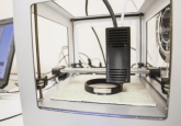
3D printing with PeptiGels®
What properties do your hydrogels have that make them suitable for 3D bioprinting?
Our peptide hydrogels are shear-thinning making them compatible with 3D bioprinting. Upon the addition of external pressure, such as passing through the needle, the gels liquify so they can move through the needle smoothly. Immediately after release from the printing nozzle/head the gels reform spontaneously.
What are differences between PeptiGels® and PeptiInks®?
PeptiGels® are hydrogels specifically designed for cell culture while PeptiInks® are bioinks specifically designed and formulated for 3D bioprinting.
What is the average percentage viability of cells within the gel when printed?
Our studies have shown that the 80–90% cells remain viable for the duration of the studies.