In focus: applications in cardiology
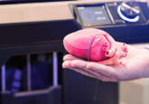
The use of 3D printing in cardiology has grown over recent years, from scientists working to fabricate cardiac tissues with bioprinting techniques to surgical teams 3D printing complex anatomy models ahead of life-changing procedures.
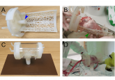
In preclinical research, for example, researchers have used 3D-printed devices to help fabricate heart cells from stem cells and regenerative patches have even been fabricated with future plans for cardiac tissue replacement and repair. Cardiac tissues have also been developed for toxicology screening and many 3D-printed devices for improving the management of cardiovascular diseases are also in the early stages of development.
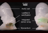
In clinical medicine, 3D printing has disrupted how surgical teams image, explain, plan and perform cardiovascular interventions, reducing the time, costs and risks associated with complex procedures, especially with patients with complex anatomies. 3D-printed models have also been used to aid machine learning programs to identify risk factors for congenital heart defects – a strategy that could be easily implemented in the near future.
In this feature, 3DMedNet consults the experts on the use of 3D printing in cardiology, from bench to bedside. Over the next few weeks, be sure to watch this space for the latest ‘Focus exclusives’ including editorials and interviews highlighting some of the latest research in this area.
As part of this feature, 3DMedNet members also gain exclusive access to relevant journal articles published in our partner publication, the Journal of 3D Printing in Medicine as well as our sister publication, Future Cardiology.
You can follow this feature via LinkedIn, Facebook and Twitter by using the hashtag, #3DIFcardiology.

